Guerbet Injector Installation Process: What to Expect During Equipment Setup

Written by: Spectrum Xray /
/
November 20, 2025

Key Takeaways

  • Guerbet injectors like OptiVantage DH (CT) and OptiStar Elite (MRI) provide precise, programmable contrast delivery for advanced imaging protocols.
  • Pre-installation checks for power, grounding, networking, room layout, and documentation are essential to avoid delays and rework.
  • Installation follows defined steps: physical setup, electrical and interface testing, fluid-path assembly, software configuration, and calibration.
  • System integration with CT/MRI consoles and radiology IT enables synchronized scanning, automated documentation, and robust contrast dose tracking.
  • Planned maintenance, periodic recalibration, and solid service contracts help keep injectors accurate, safe, and consistently available.

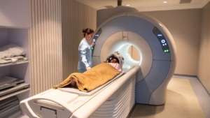
Installing a Guerbet injector in a CT or MRI suite is a structured process that goes well beyond placing new hardware in the room. Each OptiVantage or OptiStar system must be matched to facility power, network, workflow, and scanner interfaces, then calibrated to deliver contrast with high precision. By understanding the key stages—planning, delivery, setup, and verification—imaging teams can go live quickly while protecting safety, image quality, and throughput.

What Is a Guerbet Injector and How Does It Support CT and MRI Contrast Delivery?

Guerbet injectors are automated contrast delivery systems engineered for precise, controlled administration of contrast media during diagnostic imaging. These FDA-cleared devices enable technologists to program flow rates, injection volumes, and pressure limits while maintaining patient safety through built-in verification systems. Understanding the technical specifications and design features of each model is essential for successful Guerbet injector installation.

Which Guerbet Injector Models Are Typically Installed in CT and MRI Suites?

The OptiVantage DH serves CT applications with FDA 510(k) clearance obtained November 30, 2004 (K042744). This Class II device under 21 CFR 870.1650 delivers flow rates from 0.1–10.0 mL/sec with pressure limits ranging from 50–325 psi in standard configuration and up to 350 psi in multi-use systems. Syringe options include 200 mL empty syringes and prefilled sizes of 50, 75, 100, and 125 mL. Programmable delay and injection times span 0–600 seconds, accommodating diverse CT protocols.

The OptiStar Elite addresses MRI injector installation requirements with FDA clearance from May 7, 2008 (K073592). Its non-magnetic ultrasonic motor operates safely in fields up to 3 Tesla. Piezoelectric motors drive dual lead screw rams with 0.1 mL volume precision for next-generation gadolinium-based contrast agents. Battery-free operation eliminates maintenance intervals and prevents mid-scan power failures during critical examinations.

How Does Injector Design Influence Installation Requirements and Workflows?

Mounting flexibility directly impacts CT injector setup planning. Ceiling mount configurations preserve floor space in compact imaging suites, while dual-head designs enable simultaneous contrast and saline delivery without manual switching. The multi-use system permits 24-hour operation across multiple patients, accelerating workflow by 64% compared to single-patient configurations and saving an average of 35 minutes per shift.

Radiology equipment integration depends on interface capabilities built into each system. Every OptiVantage model includes CAN class 4 and relay interfacing for seamless scanner communication. The OptiStar Elite’s ceiling mount design optimizes MRI suite workflows while maintaining the non-magnetic safety profile required for high-field environments. These design considerations must inform pre-installation site assessments and injector calibration process planning.

Why Is Pre-Installation Planning Critical for a Successful Guerbet Injector Setup?

Inadequate pre-installation planning causes delays, compatibility failures, and costly rework. Successful Guerbet injector installation requires coordinated verification of electrical infrastructure, network connectivity, physical space, and regulatory compliance before equipment arrives on site. Teams that conduct thorough site assessments reduce installation time and minimize operational disruptions.

What Facility, Power, and Network Prerequisites Must Be Confirmed in Advance?

The syringe heater maintains contrast media at 37°C, requiring dedicated power circuit verification during CT injector setup. Facilities must confirm adequate amperage and proper grounding to prevent thermal system failures that compromise contrast viscosity and injection performance.

CAN class 4 and relay interfacing enable communication between the injector and scanner console. Network infrastructure assessments should verify cable routing paths, confirm interface compatibility with existing CT or MRI systems, and test signal integrity before installation day. Mismatched protocols discovered during radiology equipment integration cause preventable delays and may require hardware upgrades or adapter purchases.

How Should Room Layout, Workflow, and Patient Flow Be Considered Before Installation?

Ceiling mount versus floor mount configuration depends on available floor space and cable management requirements. Ceiling installations preserve workspace in compact suites but demand structural load verification and overhead clearance measurements. Floor-mounted systems offer repositioning flexibility but occupy valuable square footage in high-volume departments.

Dual-head system positioning must optimize technologist access to both contrast and saline syringes without obstructing patient transfer or emergency response pathways. Walk-through simulations during planning identify reach limitations, cable interference points, and sightline obstructions that compromise operator efficiency during MRI injector installation.

What Documentation, Approvals, and Stakeholder Coordination Should Be Completed Beforehand?

FDA 510(k) clearance documentation confirms Class II medical device status under 21 CFR 870.1650. Regulatory verification prevents procurement of non-compliant equipment and satisfies Joint Commission and state health department inspection requirements. Documentation should include clearance numbers K042744 for OptiVantage DH and K073592 for OptiStar Elite.

Manufacturer records identify the OptiVantage DH as originally produced by Mallinckrodt Inc., Liebel-Flarsheim Business (2111 East Galbraith Road, Cincinnati, OH 45237) under Establishment Registration 1518293. Coordinating with biomedical engineering, IT, radiology management, and clinical staff before installation ensures stakeholder alignment on workflow changes, training schedules, and injector calibration process timelines.

What Should Imaging Teams Expect on the Day of Guerbet Injector Delivery and Staging?

Delivery day transforms planning into physical reality. Installation teams coordinate equipment arrival, conduct incoming inspections, and prepare components for final positioning. Clear communication between receiving staff, biomedical engineering, and service technicians prevents delays and ensures safe handling of sensitive electronics.

How Are Shipping, Receiving, and Inspection of the Guerbet Injector Handled on Arrival?

Receiving staff verify shipment contents against packing lists and inspect external packaging for transit damage before signing delivery documentation. Damaged packaging requires photographic documentation and immediate carrier notification to protect warranty coverage. The receiving team coordinates with installation technicians to move equipment to staging areas using appropriate material handling equipment rated for the injector’s weight and dimensions.

Visual inspection confirms serial numbers match purchase orders and FDA clearance documentation. Teams check for obvious shipping damage to control interfaces, mounting hardware, and protective covers before moving units into imaging suites. Any discrepancies halt the installation process until manufacturers provide a resolution or replacement components.

How Is the Injector Unpacked, Checked, and Staged Before Installation?

The OptiVantage power head houses two electromechanical syringe drive systems requiring careful unpacking to prevent damage to drive mechanisms and electronic controls. The OptiStar Elite power head contains two piezoelectric motors with lead screw rams that demand specialized handling due to their precision engineering and MRI-compatible materials.

Installation teams inventory all components, verify accessory kits contain specified tubing and syringes, and stage items in sequence for efficient assembly. This systematic approach during CT injector setup prevents mid-installation discoveries of missing parts that extend completion timelines and delay training schedules

.

What Safety and Infection Control Measures Are Followed During Physical Setup?

Optional RFID systems enhance patient safety throughout the injector calibration process and subsequent clinical use. RFID technology prevents accidental air embolism by detecting empty or previously used syringes before injection cycles begin. The system also blocks patient cross-contamination risks by tracking syringe usage across procedures.

Medication error reduction represents another critical RFID benefit, as the system verifies contrast media type and concentration against programmed protocols. RFID integration significantly reduces syringe preparation and labeling time, streamlining workflows while maintaining sterile technique during physical setup. Installation teams should verify RFID reader function and syringe tag recognition before proceeding to radiology equipment integration steps.

What Steps Are Involved in Installing and Calibrating a New Guerbet Injector for CT or MRI Applications?

Installation progresses through mechanical positioning, electrical verification, network configuration, fluid path assembly, software setup, and calibration validation. Each step builds upon the previous phase, requiring systematic completion before advancing. Rushing through the injector calibration process creates safety risks and performance issues that compromise clinical operations.

How Is the Injector Mechanically Positioned and Secured in the CT or MRI Room?

MRI injector installation demands verification of the OptiStar Elite’s non-magnetic design for safe operation in high-field environments up to 3 Tesla. Service technicians use non-ferrous tools and confirm proper magnetic field zone positioning before securing mounting hardware. Any ferromagnetic components near the scanner bore create projectile hazards and image artifacts.

Ceiling mount installations require structural anchor verification and load testing before attaching the injector assembly. Mobile stand configurations demand wheel lock verification and stability testing to prevent movement during contrast delivery. Final positioning must allow unobstructed cable routing and maintain technologist access to control interfaces.

How Are Electrical Connections, Power Checks, and Grounding Verified?

Power system verification confirms the syringe heater reaches and maintains 37°C operating temperature. Temperature sensors and thermal feedback loops prevent overheating that degrades contrast media or creates patient safety concerns during CT injector setup. Dedicated circuit breakers rated for continuous load prevent mid-scan power interruptions.

Dual syringe drive systems require balanced power delivery to both injection heads. Technicians measure voltage stability under simulated load conditions and verify proper grounding to chassis and facility ground systems. Ground fault protection must activate within specified response times to protect patients and operators.

How Are Data, Network, and Interface Cables Configured and Tested?

CAN class 4 interface connections link the injector to scanner consoles for synchronized contrast delivery. Cable routing avoids pinch points, maintains minimum bend radii, and uses shielded pathways to prevent electromagnetic interference. Technicians verify termination resistors and check signal integrity with protocol analyzers before conducting live communication tests.

Relay interfacing capabilities enable legacy CT and MRI systems to trigger injection sequences through hardwired signals. Testing confirms proper voltage levels, verifies timing accuracy, and validates fail-safe behaviors when communication links drop. Successful radiology equipment integration requires documented verification of all interface modes before clinical use.

How Are Syringes, Tubing, and Fluid Paths Assembled and Leak-Checked?

Syringe options include 200 mL empty syringes (P/N 600096S with spike) and prefilled sizes of 50, 75, 100, and 125 mL. Tubing selections include P/N 601195 coiled extension tubing (60″L, 400 psi) and P/N 601227 with male luer adapter (48″L, 400 psi). Component selection depends on protocol requirements and distance between injector and patient access site.

Auto-fill automatically fills disposable syringes, eliminating manual aspiration and reducing air introduction. Auto purge removes residual air with a single button press, streamlining preparation sequences. The Patency Check® feature confirms proper IV placement and vascular patency before delivering contrast media, preventing extravasation injuries that delay procedures and harm patients.

How Is the Injector Software Configured with Default Protocols and User Profiles?

Dual injection protocols support saline ratios from 10% to 70% in 5% increments, enabling customized contrast dilution for diverse clinical indications. OptiBolus® software reduces contrast media usage by up to 40% without compromising image quality through intelligent delivery algorithms. This feature lowers per-scan costs and reduces patient exposure to iodinated contrast.

The Timing Bolus® feature enables test injections that confirm protocol timing before diagnostic scans begin. Technicians program operator profiles with permission levels, default protocols, and preferred interface settings. Comprehensive software configuration during the injector calibration process prevents workflow interruptions and reduces operator errors during high-volume clinical operations.

How Are Flow, Volume, and Pressure Parameters Calibrated and Verified?

OptiVantage DH calibration establishes flow rates from 0.1–10.0 mL/sec with pressure limits spanning 50–325 psi in standard configuration and 350 psi in multi-use systems. Delay times and injection durations range from 0–600 seconds, accommodating protocols from rapid bolus injections to extended infusion sequences. Calibration verification uses precision scales, pressure transducers, and timing analyzers.

OptiStar Elite MRI calibration confirms volume precision to 0.1 mL increments, essential for gadolinium-based contrast agents requiring exact dosing. Technicians validate motor response, pressure feedback loops, and safety interlocks across the full operating range. Documentation includes serial numbers, calibration dates, and technician signatures for regulatory compliance.

How Are Test Injections, Phantom Runs, and Acceptance Criteria Documented?

Test injections into collection vessels verify programmed flow rates, total volumes, and pressure profiles match specifications. The Timing Bolus® feature validates scan synchronization timing during phantom runs with simulated patient scenarios. Clinical performance benchmarks include 96% patient satisfaction rates and zero adverse events reported in recent OptiVantage studies.

Acceptance documentation records all test results, identifies any deviations from specifications, and confirms corrective actions before clinical release. Photographs capture final installation configuration, cable routing, and control interface positions. Signed acceptance forms from biomedical engineering, radiology management, and service technicians complete the Guerbet injector installation process and authorize clinical operations.

How Is a Newly Installed Guerbet Injector Integrated with Scanners and Radiology IT Systems?

Radiology equipment integration connects the injector to imaging consoles and enterprise systems for coordinated contrast delivery and data management. Communication protocols synchronize injection timing with scan acquisitions while data interfaces enable dose tracking and protocol documentation. Successful integration requires testing across multiple scanning scenarios and validating bidirectional data exchange with hospital information systems.

How Does the Injector Communicate with CT or MRI Consoles During Scanning?

The standard CAN class 4 interface provides digital communication between the injector and modern scanner consoles. This protocol enables real-time status updates, automated injection triggering, and error reporting during examinations. Technicians configure network addresses, baud rates, and message formats to match scanner specifications during CT injector setup.

Relay interfacing capabilities support legacy CT and MRI systems lacking digital communication protocols. Hardwired relay connections transmit trigger signals, ready status indicators, and completion confirmations through voltage state changes. Both interface types require validation testing across all clinical protocols to ensure reliable communication under varying load conditions and timing requirements.

How Are Protocol Triggering, Timing Signals, and Scan Synchronization Validated?

The Timing Bolus® feature confirms scan timing and synchronization by executing test injections while monitoring console communication signals. Technicians verify trigger delays match programmed values and injection sequences complete before scan acquisitions begin. Mismatched timing creates suboptimal contrast enhancement and requires recalibration of delay parameters.

Test injection validation ensures proper protocol execution across diagnostic scenarios including CT angiography, perfusion studies, and routine contrast-enhanced scans. Service teams document response times, verify emergency stop functionality propagates to scanner consoles, and confirm error conditions halt both injection and scanning. Systematic validation during MRI injector installation prevents clinical failures that compromise patient safety and image quality.

How Can Injection Data Be Linked to RIS, PACS, or Dose Management Systems?

Integration with radiology information systems enables automatic documentation of contrast type, volume, flow rate, and pressure parameters in patient records. This connectivity eliminates manual transcription errors and supports dose tracking requirements for contrast stewardship programs. Network interfaces must comply with HL7 messaging standards and DICOM structured reporting protocols.

PACS integration allows injection metadata to accompany image series for comprehensive examination documentation. Dose management systems aggregate contrast administration data across patient populations to support quality initiatives and regulatory reporting. Facilities should verify data mapping accuracy, test error handling for network interruptions, and establish backup documentation procedures for system failures during the injector calibration process.

 How Should Imaging Departments Plan for Ongoing Maintenance and Recalibration of Guerbet Injectors?

Proactive maintenance prevents unexpected failures that disrupt clinical schedules and compromise patient safety. Structured preventive maintenance programs combined with performance verification protocols ensure injectors maintain calibration accuracy throughout their service life. Strategic service arrangements minimize downtime and protect the workflow efficiencies achieved during initial Guerbet injector installation.

What Routine Checks and Preventive Maintenance Tasks Should Be Scheduled?

OptiStar Elite battery-free operation eliminates scheduled battery replacement intervals and prevents mid-scan power failures that plague battery-dependent MRI injector installation configurations. This design reduces annual maintenance costs and eliminates unplanned downtime associated with degraded battery performance. Departments avoid the expense of replacement battery modules and technician labor for battery service cycles.

The auto home feature simplifies routine ram positioning checks by automatically returning drive mechanisms to starting positions after each injection cycle. Technicians verify ram travel distances, inspect drive screw lubrication, and check alignment indicators during scheduled maintenance visits. Daily visual inspections should confirm pressure transducer readings, verify syringe heater temperatures, and test emergency stop functionality across all control interfaces.

How Often Should Performance Verification and Recalibration Be Performed?

Manufacturer recommendations typically specify annual injector calibration process cycles with interim quarterly performance checks. High-volume facilities may require semi-annual full calibrations to maintain accuracy specifications across thousands of injection cycles. Biomedical engineering departments document flow rate accuracy, pressure limit verification, and volume delivery precision using traceable measurement standards.

Triggering events beyond scheduled intervals include software updates, component replacements, or observed performance deviations during clinical use. Any pressure limit excursions, volume delivery discrepancies, or communication errors warrant immediate verification testing before returning equipment to clinical service. Documentation of all calibration activities satisfies accreditation requirements and supports regulatory inspections.

How Can Service Contracts and Support Arrangements Minimize Injector Downtime?

Multi-use systems reduce setup time and contribute to 35 minutes saved per shift by eliminating frequent syringe changes and preparation sequences. This efficiency gain translates to higher equipment utilization rates and fewer workflow disruptions requiring emergency service interventions. Departments achieve 64% faster workflows that reduce stress on mechanical components and decrease premature wear patterns.

Comprehensive service contracts provide predictable maintenance costs, priority response times, and access to factory-trained technicians familiar with CT injector setup and radiology equipment integration complexities. Contracts should include preventive maintenance visits, software updates, emergency repair coverage, and loaner equipment provisions for extended repairs. Proactive service arrangements prevent small issues from escalating into failures that halt clinical operations and force expensive expedited parts shipments.

Streamline Your Injector Setup With Spectrum Medical Imaging Co.

At Spectrum Medical Imaging Co., we make Guerbet injector installation smooth, safe, and efficient. We work with you to validate site readiness, coordinate mounting and integration, configure protocols, and support staff training so your team can use features like Timing Bolus® and OptiBolus® with confidence from day one.

We also provide ongoing maintenance and responsive technical support to keep your CT and MRI injectors calibrated, compliant, and ready for high-volume use. Reach out to us at Spectrum Medical Imaging Co. so we can help you plan and execute an injector setup that strengthens both your workflow and your patient care.

Latest Articles

Edit Post Site Icon Template Library Edit with Elementor Gadolinium-Based Contrast Agents Explained · Post Ctrl+K Save draft Publish Gadolinium-Based Contrast Agents Explained Gadolinium contrast agents represent a critical component of modern diagnostic imaging for healthcare facilities managing high-volume MRI operations. These pharmaceutical compounds enhance tissue differentiation by altering magnetic relaxation times, enabling radiologists to identify pathologies invisible on unenhanced scans. The global market reached $1.57 billion in 2025, with annual administrations estimated between 12 and 18 million procedures in the United States alone. Understanding GBCA selection directly impacts patient safety protocols, operational costs, and diagnostic accuracy across hospital systems and outpatient imaging centers. Recent regulatory guidance has shifted clinical practice toward macrocyclic agents due to superior stability profiles, while emerging low-dose formulations promise reduced gadolinium exposure without compromising image quality. This guide provides decision-makers with evidence-based analysis of chemical structures, safety comparisons, pricing tiers, and regulatory developments shaping GBCA procurement strategies. Key Takeaways Macrocyclic GBCAs provide superior stability and minimal retention compared to linear agents Neurological disorders represent 35% of the GBCA market share, followed by cardiovascular imaging at 28% Bayer AG leads the market with 28% share, followed by Bracco Imaging at 24% Mid-range tier agents cost $85-100 per dose, while premium agents range from $130-140 per dose Gadopiclenol delivers equivalent image quality at half the standard dose (0.05 mmol/kg) What Are Gadolinium-Based Contrast Agents? Gadolinium-based contrast agents are pharmaceutical compounds containing the paramagnetic metal gadolinium chelated within organic ligand structures. These agents improve MRI diagnostic accuracy by shortening T1 and T2 relaxation times in tissues where they accumulate. The chelation structure prevents free gadolinium ions from causing toxicity while maintaining the metal's magnetic properties essential for MRI enhancement. Chemical stability depends on ligand structure, binding strength, and ionic characteristics of the compound. Linear agents feature open-chain flexible ligands with weaker binding, while macrocyclic agents use cage-like rigid structures providing stronger gadolinium retention. This structural difference directly influences safety profiles, tissue retention rates, and regulatory classifications guiding clinical use. How Do GBCAs Enhance MRI Imaging? GBCAs enhance MRI imaging by shortening T1 relaxation times in tissues, creating brighter signal intensity on T1-weighted sequences. The paramagnetic properties of gadolinium create local magnetic field disturbances that accelerate hydrogen proton realignment after radiofrequency pulse excitation. This acceleration produces a stronger contrast between normal and pathological tissues, particularly in highly vascularized lesions. The enhancement effect depends on gadolinium concentration, tissue perfusion rates, and agent relaxivity characteristics. Ultra-high relaxivity agents provide 2-3 times greater signal enhancement per molecule compared to standard formulations, enabling reduced dose administration. Neuroimaging contrast applications particularly benefit from this improved tissue differentiation, revealing subtle abnormalities in brain parenchyma and vascular structures. What Are the Main Chemical Structures of GBCAs? Linear agents have open-chain flexible ligand structures that allow greater molecular movement and weaker gadolinium binding. Macrocyclic agents have cage-like, rigid ligand structures that trap gadolinium ions within stable molecular frameworks. This structural difference determines stability profiles, with macrocyclic agents demonstrating significantly lower dissociation rates in physiological conditions. Ionic agents have higher osmolality and higher stability compared to nonionic formulations. Nonionic agents have lower osmolality and more comfortable injection profiles, reducing adverse reactions at the injection site. Ultra-high relaxivity agents provide 2-3 times higher relaxivity than standard formulations, enabling diagnostic image quality at half the conventional dose. Why Are GBCAs Used in Clinical Practice? GBCAs are used in clinical practice to detect pathologies invisible on unenhanced MRI scans, including tumors, inflammation, and vascular abnormalities. These agents enable radiologists to characterize lesion margins, assess tissue perfusion, and differentiate benign from malignant processes. Approximately 30-45% of the 40 million annual MRI procedures in the United States utilize contrast enhancement for improved diagnostic accuracy. The clinical decision to administer contrast depends on the anatomical region, suspected pathology, and patient risk factors. High-risk populations, including those with renal impairment, require careful agent selection to minimize adverse outcomes. The critical role of CT and MRI contrast media in patient diagnosis extends across multiple specialties, from oncology to neurology. Which Medical Conditions Benefit Most from GBCAs? Neurological disorders represent 35% of the GBCA market share, driven by brain tumor imaging, stroke evaluation, and multiple sclerosis monitoring. Cardiovascular imaging accounts for 28% market share, including myocardial perfusion studies and coronary artery assessments. Cancer imaging represents 18% market share, with applications in tumor staging, treatment response evaluation, and metastasis detection. Musculoskeletal disorders comprise 10% market share, focused on joint abnormalities, soft tissue masses, and spinal pathology. Abdominal imaging accounts for 6% market share, primarily for liver lesion characterization and pancreatic disease evaluation. The remaining 3% includes specialized applications across various organ systems requiring enhanced tissue differentiation. How Are GBCAs Utilized Among Different Patient Groups? Annual MRI procedures in the United States reach approximately 40 million examinations across all patient populations. The percentage using GBCAs ranges from 30-45%, depending on clinical indication and institutional protocols. Estimated annual GBCA administrations total 12-18 million doses, representing substantial pharmaceutical utilization within diagnostic imaging operations. Geriatric populations demonstrate the highest utilization rates due to increased chronic disease prevalence and oncologic surveillance needs. Pediatric applications are increasing, with a strong preference for macrocyclic agents to minimize long-term retention risks. Safety-first approaches to managing contrast allergies in high-risk patients require specialized protocols for those with previous adverse reactions or compromised renal function. What Are the Key Differences Between Linear and Macrocyclic GBCAs? Linear and macrocyclic GBCAs differ fundamentally in molecular structure, binding stability, and safety profiles during clinical use. Linear agents feature open-chain ligands with moderate binding strength, allowing greater gadolinium dissociation in physiological environments. Macrocyclic agents employ rigid cage-like structures that trap gadolinium ions within stable molecular frameworks, preventing release. These structural differences translate into distinct clinical safety profiles and regulatory classifications guiding agent selection. Market trends show declining linear agent use as institutions transition toward macrocyclic formulations to reduce liability exposure. The role of iodine-based and gadolinium-based agents in diagnostics requires understanding both agent classes for comprehensive imaging protocols. How Do Stability and Safety Profiles Compare? Linear agents have weak to moderate binding strength, while macrocyclic agents have very strong binding strength that resists dissociation. Linear agents have lower stability in physiological conditions versus macrocyclic agents, which maintain high stability across temperature and pH ranges. Linear agents carry high NSF risk (Group I classification) while macrocyclic agents demonstrate very low NSF risk (Group II classification). Linear agents show high tissue retention patterns, including brain deposition, while macrocyclic agents exhibit minimal retention with faster clearance. Linear agents are declining or phasing out of institutional formularies versus macrocyclic agents, which are dominant and growing. Gadolinium MRI contrast safety for patients with kidney disease requires exclusive use of macrocyclic formulations in renal-impaired populations. What Are Examples of Common Linear and Macrocyclic Agents? Among linear agents, Magnevist (gadopentetate dimeglumine) by Bayer carries a Group I NSF classification under ACR guidelines—the highest-risk category—and is contraindicated for patients with renal impairment. MultiHance (gadobenate dimeglumine) by Bracco is an ionic linear agent with higher relaxivity than standard linear formulations; however, like Magnevist, it is subject to institutional phase-out as facilities transition to macrocyclic protocols. Macrocyclic agents include Gadavist (gadobutrol) by Bayer, classified as Group II under ACR guidelines and used widely across CNS imaging. Dotarem (gadoterate meglumine) by Guerbet maintains an established Group II safety profile, frequently selected for patients with borderline renal function. ProHance (gadoteridol) by Bracco is used across CNS, spine, and body MRI applications with similar safety characteristics. Elucirem (gadopiclenol) by Guerbet features high relaxivity, delivering equivalent diagnostic image quality at a lower administered dose of 0.05 mmol/kg. Vueway (gadopiclenol) by Bracco represents a newer macrocyclic GBCA with provisional Group III status pending long-term real-world safety data. Bayer brands include both Gadavist (gadobutrol) for general imaging and Eovist (gadoxetate) for specialized liver applications. How Do Pricing and Market Competition Shape GBCA Use? Pricing and market competition directly influence institutional formulary decisions, affecting which agents remain available for routine clinical use. Mid-range tier agents priced at $85-100 per dose represent the current standard of care across most healthcare systems. Premium tier agents commanding $130-140 per dose target specialized applications, including low-dose protocols and organ-specific imaging. Budget constraints force procurement teams to balance cost containment with safety profile preferences and regulatory compliance requirements. Single-source vendor consolidation through suppliers like Spectrum Medical Imaging Co. can reduce administrative burden while ensuring competitive pricing. Comparative analyses such as Gadobutrol vs Dotarem help decision-makers evaluate equivalent agents within similar price tiers. Which Companies Lead the GBCA Market? Bayer AG holds 28% market share, leading through Gadavist and Eovist brands with strong institutional penetration. Bracco Imaging commands 24% market share, competing through ProHance and the recently launched Vueway formulation. GE Healthcare maintains 22% market share despite regulatory challenges affecting linear agent portfolios. Guerbet controls 16% market share, driven by Dotarem's established safety record and Elucirem's innovative low-dose profile. Lantheus Medical Imaging holds 6% market share, focusing on specialized cardiovascular imaging applications. The remaining 4% comprises smaller manufacturers and regional distributors serving niche market segments. How Are Agents Categorized by Price and Application? Mid-range tier agents include ProHance (gadoteridol), Gadavist (gadobutrol), and Dotarem (gadoterate), priced at $85-100 per dose. These formulations represent the standard of care for routine MRI enhancement across most clinical indications. Gadobutrol from Fresenius-Kabi offers a generic alternative within this mid-range category, providing comparable clinical performance for facilities managing formulary budgets without sacrificing macrocyclic safety profiles. Budget tier pricing at $45-50 per dose applies to legacy linear agents declining in use due to safety concerns. Premium tier agents include Elucirem/Vueway (gadopiclenol) and Eovist (gadoxetate), commanding $130-140+ per dose for specialized applications. The premium tier targets low-dose protocols, organ-specific imaging, and high-risk populations requiring maximum safety margins. Dotarem's specific advantages in improving MRI imaging outcomes justify premium pricing in certain clinical contexts despite higher per-dose costs. What Factors Influence Radiologists' Choices of GBCAs? Radiologists' GBCA selection depends on multiple factors, including patient safety profiles, diagnostic image quality requirements, and institutional availability constraints. Patient safety ranks as the top priority for 93% of radiologists surveyed, reflecting increased awareness of retention and NSF risks. Image quality considerations influence 92% of radiologists' decisions, as diagnostic confidence depends on adequate tissue enhancement. Supply availability concerns affect 87% of radiologists, particularly following manufacturer shortages and distribution disruptions in recent years. Cost effectiveness influences 72% of radiologists' decisions, though safety considerations typically override price concerns for high-risk populations. Gadolinium retention concerns affect 86% of radiologists, driving preference shifts toward macrocyclic agents and low-dose formulations. How Important Are Safety and Image Quality? Patient safety is prioritized by 93% of radiologists when selecting contrast agents for clinical use. Image quality ranks nearly equal at 92%, as inadequate enhancement compromises diagnostic accuracy and may require repeat examinations. Supply availability concerns 87% of radiologists, reflecting recent market disruptions affecting workflow continuity. Cost effectiveness influences 72% of radiologists' decisions, though typically as a secondary consideration after safety and quality. Gadolinium retention concerns affect 86% of radiologists, particularly when imaging pediatric patients or those requiring serial examinations. Considerations such as whether contrast media should be warmed before injection demonstrate attention to patient comfort alongside safety priorities. What Are Radiologists' Views on Low-Dose and Environmental Concerns? Reduced gadolinium dose interests 99% of radiologists surveyed, with 60% expressing very strong interest in low-dose formulations. Somewhat interested radiologists comprise 39%, indicating near-universal support for dose reduction strategies when diagnostic quality remains equivalent. Supply availability concerns persist among 55% of radiologists, driving dual-source procurement strategies at many institutions. NSF risk concerns affect 28% of radiologists, concentrated among those serving renal-impaired populations requiring specialized protocols. Environmental concerns regarding gadolinium contamination influence 35% of radiologists, though this remains secondary to patient-focused safety priorities. Cost concerns affect 42% of radiologists, particularly in resource-constrained settings, balancing quality with budgetary limitations. How Are Regulatory Bodies Guiding GBCA Usage? Regulatory bodies, including the FDA and European Medicines Agency, have issued specific guidance restricting linear agent use in high-risk populations. These regulations mandate macrocyclic agent preference for patients with renal impairment, pregnant women, and pediatric populations. Mandatory medication guides and enhanced pharmacovigilance requirements now apply to all GBCA formulations regardless of chemical structure. Recent approvals demonstrate regulatory support for low-dose innovations that reduce total gadolinium exposure while maintaining diagnostic accuracy. Institutions must implement policies aligning with ACR and ESUR guidelines to minimize liability exposure and ensure regulatory compliance. Regulatory oversight continues evolving as long-term retention data accumulates across different agent classes and patient populations. What Are Recent Approvals and Restrictions? February 2026 brought expanded gadopiclenol approval for neonates and infants under 2 years, enabling low-dose protocols in vulnerable pediatric populations. November 2025 marked Bracco's delivery of 3 million VUEWAY doses, demonstrating a successful commercial launch and market acceptance. August 2025 saw Bayer's Gadoquatrane NDA accepted for FDA review, potentially introducing another ultra-low-dose option. October 2023 brought European Commission approval for Elucirem (gadopiclenol), establishing a regulatory precedent for high-relaxivity low-dose formulations. FDA warnings on gadolinium retention in brain tissue have driven institutional policy changes favoring macrocyclic agents. These regulatory actions reflect growing emphasis on minimizing cumulative gadolinium exposure across patient lifetimes. How Do Guidelines Affect High-Risk Populations? Linear agents are contraindicated for patients with renal impairment due to elevated NSF risk profiles. Group II macrocyclic agents are exclusively recommended for patients with renal impairment, reflecting superior safety characteristics. Macrocyclic agents are strongly preferred for pregnant women to minimize fetal gadolinium exposure during gestation. Mandatory medication guides are required for all GBCA formulations to inform patients of potential retention risks. Enhanced pharmacovigilance programs monitor long-term outcomes across different agent classes and patient populations. Renal safety protocols require pre-procedure creatinine assessment and eGFR calculation to identify high-risk patients requiring specialized agent selection. What Emerging Technologies Are Impacting GBCA Development? Emerging technologies focus on reducing gadolinium dose requirements through improved relaxivity characteristics and alternative paramagnetic metals. Ultra-high relaxivity formulations enable equivalent diagnostic quality at half the standard dose, reducing total gadolinium burden. Novel chemical structures provide enhanced tissue specificity, improving lesion detection while minimizing background enhancement. Artificial intelligence integration optimizes contrast timing and dose calculations based on patient-specific factors, including body weight and renal function. These technological advances address radiologists' demand for safer, more efficient contrast protocols without compromising diagnostic accuracy. Market growth projections reaching $10.8 billion by 2030 reflect continued innovation investment across major manufacturers. Which Innovations Aim to Reduce Gadolinium Dose? Gadoquatrane by Bayer is currently under FDA review as a potential ultra-low-dose macrocyclic formulation. This compound targets 50% dose reduction compared to standard agents while maintaining equivalent diagnostic image quality. Early clinical data demonstrate non-inferior performance at 0.05 mmol/kg versus conventional 0.1 mmol/kg dosing. Gadopiclenol (marketed as Elucirem and Vueway) already provides 2-3 times higher relaxivity than standard formulations, enabling half-dose protocols. These high-relaxivity agents address the 99% of radiologists interested in reduced gadolinium dose options. Clinical adoption depends on regulatory approvals, institutional formulary decisions, and long-term safety data from post-market surveillance programs. Are There Alternatives to Gadolinium Agents? Manganese-based agents are currently in GE Healthcare Phase I trials as of 2024, representing the first major alternative to gadolinium. These compounds utilize manganese's paramagnetic properties to achieve MRI enhancement without gadolinium exposure. Early preclinical data suggest comparable relaxivity characteristics with potentially different biodistribution and elimination pathways. Iron oxide nanoparticles represent another research direction, though clinical development remains in early stages. These alternatives face substantial regulatory hurdles requiring comprehensive safety and efficacy data before clinical approval. Success would fundamentally reshape the contrast agent market by eliminating gadolinium retention concerns entirely. The Current Landscape and Future Outlook for Gadolinium-Based Contrast Agents The global CT and MRI contrast agents market reached $6.22 billion USD in 2024, with MRI-specific agents representing $1.57-1.69 billion USD. Market projections show a CAGR of 7.81% from 2025-2030, reaching $10.8 billion USD by 2030. North America dominates with $2.4 billion, representing 34% of the global market, driven by advanced imaging infrastructure and high procedure volumes. Macrocyclic agent dominance continues growing as institutions phase out linear formulations in response to regulatory guidance and retention concerns. Low-dose innovations, including gadopiclenol, represent the next evolution, addressing radiologists' demand for reduced gadolinium exposure without compromising diagnostic accuracy. Market competition among Bayer, Bracco, Guerbet, and GE Healthcare drives continued innovation investment in safety and efficacy improvements. Healthcare facilities must balance safety priorities, cost constraints, and supply chain reliability when establishing GBCA procurement strategies. Dual-sourcing arrangements with reliable suppliers like Spectrum Medical Imaging Co. help ensure uninterrupted contrast availability during manufacturer shortages. Institutions implementing macrocyclic-only formularies position themselves favorably in an increasingly safety-conscious regulatory environment while maintaining diagnostic capabilities across all clinical indications. Source GBCAs with Confidence - Partner with Spectrum Medical Imaging Co. With over 30 years of experience supplying hospitals, imaging centers, and outpatient facilities across the West Coast, Spectrum Medical Imaging Co. delivers the right GBCAs — from macrocyclic agents like Gadavist, Dotarem, ProHance, and Elucirem to specialized formulations like Eovist — at competitive pricing your budget can rely on. We combine guaranteed supply chain reliability with expert guidance on formulary transitions, helping your team align contrast procurement with current ACR safety guidelines and regulatory requirements. Contact us to speak with a contrast media specialist and build a GBCA procurement strategy that keeps your imaging operations running without interruption. Move upMove downToggle panel: Yoast SEO SEO Readability Schema Social Focus keyphraseHelp on choosing the perfect focus keyphrase(Opens in a new browser tab) CT and MRI contrast agents Get related keyphrases(Opens in a new browser tab) Search appearance Determine how your post should look in the search results. Google preview Mobile Google preview: Switch to desktop preview. Currently showing mobile preview. Desktop Url preview: Spectrum Medical X-Ray Co spectrumxray.com SEO title preview: Gadolinium-Based Contrast Agents: Uses, Safety Guide Meta description preview: Apr 8, 2026 - Gadolinium-based contrast agents enhance MRI imaging through specific magnetic properties. Learn about GBCA types, safety profiles, pricing, and ... SEO title Insert variable Generate SEO title Gadolinium-Based Contrast Agents: Uses, Safety Guide Slug gadolinium-based-contrast-agents-explained Meta description Insert variable Generate meta description Gadolinium-based contrast agents enhance MRI imaging through specific magnetic properties. Learn about GBCA types, safety profiles, pricing, and regulatory guidance for clinical decision-making. SEO analysisOK CT and MRI contrast agents Add related keyphrase Premium Track SEO performance Content blocks New Internal linking suggestions Premium Cornerstone content Advanced Insights Move upMove downToggle panel: Glossary Post Override Disable Glossary for this post checkedunchecked Select specific glossary categories checkedunchecked Brands & Partnerships checkedunchecked Clinical & Application Training checkedunchecked Contrast Media & Consumables checkedunchecked Customer Support & Technical Expertise checkedunchecked Imaging Equipment & Technology checkedunchecked Key Decision Makers checkedunchecked Medical Equipment Maintenance & Services checkedunchecked Procurement & Logistics checkedunchecked Project Management & Planning checkedunchecked Regulatory Compliance & Safety By selecting one or more categories, only terms belonging to these will be linked Move upMove downToggle panel: Blog Post Block Gadolinium-Based Contrast Agents Explained The current image has no alternative text. The file name is: Gadolinium-Based-Contrast-Agents-Explained.jpg Replace Remove Generate with Elementor AI Add an excerpt… 2,827 words, 15 minutes read time. Last edited a second ago. Status Draft Publish April 8, 2026 9:07 am UTC+0 Slug gadolinium-based-contrast-agents-explained Author (No author) Template Default template Revisions 2 Move to trash Yoast SEO SEO analysis: OK Readability analysis: Good Improve your post with Yoast SEO Categories Search Categories Search Categories Blog Contrast Injector Repair and Maintenance Contrast Injectors Contrast Media Imaging Equipment Imaging Equipment Repair and Maintenance Radiation Shielding Services Radiographic Room and Design Planning Add Category Tags Add Tag CT and MRI contrast agents (1 of 1)CT and MRI contrast agents Separate with commas or the Enter key. Most Used imaging equipmentcontrast media injectorContrast mediamedical imaging equipment and agents distributorContrast Power InjectorsVieworks DR panels Los Angelescontrast injector serviceDR panelsmedical imaging equipmentdigital radiography (DR) panels Move upMove downToggle panel: LLMS Amplifier Exclude from llms.txt files When checked, this content will not appear in any generated llms.txt or llms-full.txt files, regardless of SEO plugin settings. Open publish panel Post NotificationsDraft saved. Close dialog Featured image Upload filesStock PhotosMedia Library Expand Details Filter mediaFilter by type Images Filter by date All dates Smush: All images Search media Media list Showing 1 of 1 media items Load more Attachment Details Gadolinium-Based-Contrast-Agents-Explained.jpg April 13, 2026 38 KB 1280 by 720 pixels Edit with Elementor AI Edit Image Delete permanently Alt Text Learn how to describe the purpose of the image(opens in a new tab). Leave empty if the image is purely decorative.Title Gadolinium-Based Contrast Agents Explained Caption Description File URL: https://spectrumxray.com/wp-content/uploads/2026/04/Gadolinium-Based-Contrast-Agents-Explained.jpg Copy URL to clipboard Smush 7 images reduced by 41.3 KB (14.5%) Main Image size: 38.29 KB Restore original Required fields are marked * Regenerate Thumbnails Regenerate Thumbnails Selected media actionsSet featured image No file chosen Select Files
Blog
Spectrum Xray

Gadolinium-Based Contrast Agents Explained

Gadolinium contrast agents represent a critical component of modern diagnostic imaging for healthcare facilities managing high-volume MRI operations. These pharmaceutical compounds enhance tissue differentiation by

Read More »