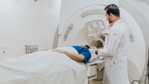
MRI contrast agents and CT contrast agents differ fundamentally in how they enhance diagnostic images. Gadolinium-based compounds alter the magnetic properties of nearby tissues, while iodinated substances physically block X-rays to create visible contrast. These distinct mechanisms affect safety profiles and clinical applications, making the choice between imaging modality differences critical for accurate diagnosis. Understanding contrast type characteristics helps healthcare facilities optimize patient outcomes while managing costs and safety risks.
Key Takeaways
- Gadolinium-based agents enhance MRI signals without ionizing radiation, while iodinated agents block X-rays in CT scans
- Allergic reactions occur less frequently with gadolinium (1 in 170,000) compared to iodine (1 in 100,000)
- Contrast-induced nephropathy affects 20-40% of patients with severe kidney disease receiving iodinated agents
- Group II macrocyclic gadolinium agents reduce nephrogenic systemic fibrosis risk to less than 0.07%
- Patient factors, including kidney function, age, and prior allergic reactions, determine optimal contrast agent selection
What Are MRI and CT Contrast Agents?
MRI contrast agents and CT contrast agents are specialized substances that improve the visualization of internal structures during diagnostic imaging. These agents work through different physical mechanisms based on the radiologic imaging technology used. MRI agents modify how tissues respond to magnetic fields, while CT agents change how X-rays pass through the body.
How Do MRI Contrast Agents Work?
Gadolinium agents enhance proton relaxivity in a magnetic field by shortening T1 and T2 relaxation times. This mechanism provides superior soft tissue contrast without exposing patients to ionizing radiation. Gadolinium chelates remain in vascular and interstitial spaces, allowing detailed visualization of blood vessels and tissue perfusion, making MRI contrast agents ideal for neurological and musculoskeletal examinations.
Gadolinium provides no ionizing radiation vs. iodine involves radiation exposure during CT procedures. This fundamental difference influences clinical decision-making when patient radiation exposure must be minimized. A complete guide to contrast agents explains the chemical composition and binding mechanisms, ensuring gadolinium safety. The absence of radiation makes gadolinium preferable for pediatric patients and repeated imaging studies.
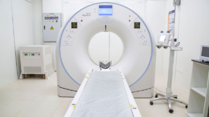
How Do CT Contrast Agents Work?
Positive contrast agents increase X-ray attenuation by absorbing photons more effectively than surrounding tissues. Iodine’s high atomic number creates strong attenuation, appearing bright on CT images, enabling clear visualization of blood vessels, organs, and pathological processes. Enhancement degree depends on iodine concentration and tissue perfusion rates.
Negative contrast agents decrease X-ray attenuation by containing low-density substances like air or gas, appearing dark on CT images. Air and carbon dioxide gas are negative contrast agents commonly used in CT colonography. Radiopaque substances absorb X-rays more readily than surrounding tissues, creating clear anatomical delineation. CT contrast agents block X-rays vs. MRI agents alter magnetic properties, representing fundamentally different physical interactions.
What Are the Key Differences Between MRI and CT Contrast Agents?
The primary differences lie in the mechanism of action, safety profiles, and clinical applications. MRI agents modify tissue magnetization without ionizing radiation, while CT agents physically block X-ray photons. These distinctions affect patient selection criteria, risk assessment protocols, and imaging study appropriateness. Cost considerations also vary significantly, with gadolinium agents costing 4 to 5 times more than iodinated compounds.
What Are Their Mechanisms of Action?
Gadolinium compounds function by altering the magnetic relaxation properties of nearby hydrogen protons. The paramagnetic gadolinium ion shortens T1 relaxation time, producing a bright signal on T1-weighted sequences, enhancing visualization of vascular structures and tissue perfusion. The mechanism depends entirely on magnetic field interactions rather than radiation absorption.
Iodinated agents work through X-ray attenuation based on iodine’s electron density and atomic number. X-ray photons interact with iodine atoms through photoelectric absorption and Compton scattering, reducing the number of photons reaching the detector and creating contrast. The effect is immediate and dose-dependent, with higher iodine concentrations producing greater attenuation.
How Do Their Safety Profiles Compare?
Severe allergic reactions occur in approximately 1 in 100,000 patients with iodine-based agents. Gadolinium allergic reactions less than 1 in 170,000, making them statistically safer for hypersensitivity concerns. NSF risk from newer Group II macrocyclic GBCAs is less than 0.07% even in patients with advanced kidney disease. Higher risk of systemic reactions with iodinated agents compared to barium requires careful patient screening.
Risk of contrast-induced nephropathy with iodinated agents affects 20-40% of patients with eGFR below 30 ml/min. Managing contrast media extravasation becomes critical when administering high-volume iodinated injections. Five percent could experience permanent renal damage with eGFR below 30 ml/min receiving iodinated contrast. Group II GBCAs are recommended over Group I for severe kidney disease patients to minimize nephrogenic systemic fibrosis risk.
What Are Their Typical Clinical Applications?
Iodinated compounds used in angiography, CT scans, and gastrointestinal examinations provide rapid vascular and anatomical assessment. CT angiography employs iodinated agents to visualize coronary arteries, pulmonary emboli, and aortic dissections. CT colonography uses gas contrast agents to distend the colon without liquid contrast. Gastrografin (diatrizoate meglumine and diatrizoate sodium) is a water-soluble oral and rectal iodinated contrast used in gastrointestinal fluoroscopy and CT when barium is contraindicated, such as in suspected bowel perforation. Emergency trauma protocols rely heavily on iodinated CT contrast for rapid hemorrhage detection.
Gadolinium agents excel in brain tumor characterization, spinal cord imaging, and musculoskeletal soft tissue evaluation. Contrast-enhanced MRI provides superior detection of multiple sclerosis lesions and subtle brain abnormalities. Cardiac MRI with gadolinium assesses myocardial viability and perfusion without radiation exposure. The choice between imaging modalities depends on the diagnostic question, tissue type, and patient-specific risk factors.
Which Types of Contrast Agents Are Used in MRI and CT?
Multiple contrast agent formulations exist within each category, with distinct chemical structures and safety profiles. Gadolinium-based agents include macrocyclic and linear formulations with varying stability characteristics. Iodinated agents range from ionic to non-ionic compounds with different osmolality properties. Additional categories include barium suspensions and microbubble agents for specialized applications.
What Are Gadolinium-Based Contrast Agents?
GBCAs are gadolinium-based contrast agents that chelate toxic gadolinium ions within stable molecular structures. Group II macrocyclic GBCAs are newer gadolinium agents with enhanced stability, reducing free gadolinium release. These formulations demonstrate superior safety profiles in patients with compromised renal function.
Gadavist (gadobutrol) by Bayer features macrocyclic GBCA with Group II classification under ACR guidelines, used widely across CNS, body, and vascular MRI protocols due to high relaxivity. Dotarem (gadoterate meglumine) by Guerbet represents a macrocyclic GBCA with an established Group II safety profile. ProHance (gadoteridol) by Bracco is a macrocyclic Group II GBCA widely used for CNS, spine, and body MRI due to its favorable safety record in renally impaired patients. MultiHance (gadobenate dimeglumine) by Bracco provides enhanced protein binding for improved vascular and hepatobiliary imaging. Gadobutrol by Fresenius-Kabi is a cost-effective macrocyclic alternative for formularies managing contrast media budgets. Eovist (gadoxetate disodium) by Bayer is a hepatocyte-specific agent used for liver lesion detection and characterization, offering both dynamic vascular phase and hepatobiliary phase imaging. Understanding Dotarem’s benefits reveals why it is frequently selected for patients with borderline renal function.
What Are Iodinated Contrast Agents?
Omnipaque (iohexol) by GE HealthCare represents the most widely used low-osmolar nonionic iodinated agent, spanning CT body, vascular, and oncology protocols with a proven safety record. Isovue (iopamidol) by Bracco and Optiray (ioversol) by Guerbet are widely used low-osmolar nonionic alternatives for CT vascular, body, and oncology protocols, offering comparable tolerability and safety profiles. Cysto-Conray (iothalamate meglumine) by Guerbet serves as a dedicated agent for retrograde cystography and urological imaging procedures. Iodine-based agents are water-soluble vs. barium is an insoluble suspension, enabling intravenous administration. Modern non-ionic formulations reduce adverse reaction rates compared to older ionic compounds.
Low-osmolar and iso-osmolar iodinated agents minimize osmotic tissue damage during injection, reducing pain at injection sites and decreasing contrast-induced adverse events. Visipaque (iodixanol) by GE HealthCare is the leading iso-osmolar iodinated agent, providing the closest osmolality match to blood and offering the highest tolerability for high-risk patients, including those with renal compromise or cardiovascular disease. Solutions to overcome iodinated contrast media shortages address supply chain challenges affecting healthcare facilities. Non-ionic agents demonstrate better tolerability profiles than older ionic formulations.
How Do Barium and Microbubble Agents Fit In?
Barium sulfate used in gastrointestinal examinations provides excellent mucosal coating for upper GI studies. Specific portfolio formulations include E-Z-HD, a high-density barium sulfate suspension for detailed mucosal coating in esophageal and upper GI studies; Readi-Cat (barium sulfate suspension) used in CT abdominal and pelvic protocols; Breeza (barium sulfate suspension) formulated for CT enterography with improved palatability; and Genus Citra by Genus, used in upper GI and small bowel fluoroscopic examinations. Barium cannot be injected into the bloodstream vs. iodine can be administered intravenously for vascular imaging. Barium’s high atomic number creates superior radiopacity for esophageal, gastric, and colonic evaluation without systemic absorption.
Definity (perflutren lipid microspheres) by Lantheus Medical Imaging serves as a primary cardiac and left ventricular opacification CEUS agent. Lumason (sulfur hexafluoride lipid-type A microspheres) by Bracco received FDA approval for liver lesion characterization in adult and pediatric patients. Microbubbles have a short half-life, typically clearing from circulation within minutes, and require specialized ultrasound equipment capable of detecting harmonic resonance frequencies.
How Do Patient Factors Influence Contrast Agent Choice?
Patient-specific variables significantly impact contrast agent selection and dosing protocols. Renal function, age, prior allergic reactions, and pregnancy status determine appropriate agent selection. Comprehensive pre-procedure risk assessment identifies patients requiring alternative imaging strategies or prophylactic medications. Balancing safety and clarity in contrast media use requires systematic evaluation of individual risk factors.
What Are the Risks for Patients with Kidney Disease?
Twenty to forty percent of patients with eGFR below 30 ml/min may develop CIN following iodinated contrast administration. Five percent could experience permanent renal damage requiring dialysis initiation. NSF risk with older linear GBCAs in patients with eGFR below 15 ml/min prompted the development of safer macrocyclic alternatives. Magnevist (gadopentetate dimeglumine) by Bayer is a Group I linear GBCA whose use is now restricted in patients with severe renal impairment due to elevated NSF risk — macrocyclic alternatives are strongly preferred in this population. Group II GBCAs are recommended over Group I for severe kidney disease patients to minimize gadolinium deposition concerns.
Gadolinium MRI contrast safety for patients with kidney disease requires careful eGFR assessment before administration. Risk of permanent renal damage in patients with eGFR below 30 ml/min necessitates hydration protocols and dose minimization strategies. Alternative imaging without contrast should be considered when the diagnostic benefit does not outweigh the nephrotoxicity risk. Dialysis patients can safely receive Group II macrocyclic agents with post-procedure dialysis within hours.
How Does Age Affect Contrast Agent Selection?
Children account for 4% of imaging examinations, requiring weight-based dosing and radiation minimization. Adults represent 66% of imaging examinations, with standard contrast protocols typically applied. Older Adults account for 30% of imaging examinations, facing increased risk from age-related renal decline. Clinical guidelines recommend reducing the contrast dose by at least 10% for elderly patients.
Pediatric contrast administration requires careful calculation based on body weight rather than standard adult volumes. Age-related decline in glomerular filtration rate increases elderly patients’ susceptibility to contrast-induced nephropathy. Geriatric patients often take multiple nephrotoxic medications that compound the risk of contrast-related renal injury. Pre-procedure hydration becomes especially important in elderly patients with marginal renal reserve.
What Are Patient Preferences and Comfort Considerations?
Patient Comfort rated 65% importance in contrast agent selection according to recent surveys. Injection site pain, warmth sensation, and metallic taste influence patient experience during contrast administration. Warming contrast media to body temperature reduces viscosity and improves injection comfort. Volume of contrast administered affects patient tolerance, with smaller volumes preferred when diagnostically adequate.
Previous adverse reactions to specific contrast agents require alternative formulation selection or premedication protocols. Patient anxiety about contrast administration can be reduced through a clear explanation of expected sensations. Some patients prefer oral contrast preparations over intravenous administration when both provide adequate diagnostic information.
What Advances Are Emerging in Contrast Agent Technology?
Novel contrast agents and dose optimization strategies are transforming diagnostic imaging safety profiles. Manganese-based compounds offer potential alternatives to gadolinium with different elimination pathways. High-relaxivity formulations enable diagnostic imaging at substantially reduced gadolinium doses. Artificial intelligence tools calculate patient-specific minimum effective doses based on clinical indications.
What Are Novel MRI Agents like Manganese-Based Compounds?
Manganese-based MRI contrast agent by GE HealthCare completed Phase I trials showing good tolerability without serious adverse events. This first-of-its-kind macrocyclic manganese agent provides an alternative mechanism, avoiding gadolinium exposure concerns through hepatobiliary excretion rather than renal clearance. Early clinical data suggest comparable enhancement characteristics to gadolinium-based formulations.
Elucirem (gadopiclenol) by Guerbet, a novel macrocyclic GBCA, received regulatory approval based on superior stability and lower required dosing. This high-relaxivity agent achieves diagnostic image quality at reduced gadolinium concentrations. Clinical trials demonstrate non-inferiority to standard-dose agents while minimizing total gadolinium exposure, addressing patient and regulatory concerns about gadolinium accumulation in brain tissues.
How Is Dose Optimization Improving Safety?
Gadopiclenol provides comparable efficacy at up to 60% lower dose of gadolinium compared to standard agents, directly addressing safety concerns while maintaining diagnostic image quality. Utilize AI and machine learning tools to calculate the minimum effective dose based on patient anatomy and scanning parameters. Advanced algorithms analyze prior imaging data to predict optimal contrast timing and volume.
Calculate dose based on patient weight and clinical indications rather than applying standardized protocols universally. Individualized dosing reduces unnecessary contrast exposure while ensuring adequate enhancement. Optimal contrast flow rates for different CT studies demonstrate how protocol customization improves both safety and image quality. Weight-based calculations prevent underdosing in larger patients and overdosing in smaller individuals.
What Are Recent Developments in Oral CT Contrast Agents?
Vueway by Bracco represents a new dark oral contrast agent improving bowel visualization on CT. Bracco delivered 3 million VUEWAY doses, meeting substantial clinical demand. Clinical Phase 2 results showed the agent revealed previously undetectable findings, including very small bowel tumors, providing superior mucosal coating compared to traditional positive oral contrast agents.
Dark oral contrast creates negative luminal contrast that improves the detection of enhancing bowel wall abnormalities, simplifying interpretation by eliminating beam-hardening artifacts. Enhanced mucosal visualization enables earlier detection of inflammatory bowel disease and small neoplastic lesions with good patient tolerability and minimal gastrointestinal side effects.
How Do Market Trends Impact Contrast Agent Availability and Use?
Global market dynamics influence contrast agent availability, pricing, and clinical protocol development. Regional adoption patterns reflect infrastructure investment, healthcare spending, and regulatory environments. Supply chain disruptions periodically force facilities to modify imaging protocols and agent selection. Cost considerations increasingly drive formulary decisions as healthcare systems manage budget pressures.
Which Regions Lead in Contrast Agent Adoption?
North America holds 38.92% revenue share in 2025, driven by advanced healthcare infrastructure and high imaging volumes, demonstrating the highest adoption rates of premium gadolinium agents and specialized formulations. Asia Pacific’s projected CAGR of 9.5% represents the fastest-growing market, fueled by expanding healthcare access. Emerging markets increasingly invest in modern imaging equipment requiring contemporary contrast agent formulations.
Urban centers use more premium agents vs. rural facilities use standard agents due to budget and equipment constraints. Metropolitan hospitals with advanced MRI capabilities more frequently employ high-relaxivity gadolinium formulations. Rural facilities often rely on standard iodinated agents and basic barium preparations due to cost considerations. The critical role of CT and MRI contrast media extends across all healthcare settings, regardless of geographic location.
How Does Cost Influence Contrast Agent Selection?
Gadolinium agents cost 4 to 5 times more than iodinated compounds, significantly impacting formulary decisions. Cost rated 72% importance in healthcare facility contrast agent selection according to procurement surveys. Budget constraints drive facilities toward generic formulations when clinically appropriate. High-volume imaging centers negotiate bulk purchasing agreements to reduce per-dose costs.
Supply shortages of iodinated contrast forced many facilities to implement conservation protocols during recent disruptions. Alternative imaging strategies without contrast became necessary when supply could not meet clinical demand. Facilities stockpile critical contrast agents to maintain operational continuity during supply chain interruptions.
What Are Expert Guidelines for Safe Contrast Agent Use?
Professional organizations publish comprehensive guidelines addressing contrast agent selection, dosing, and monitoring protocols. The American College of Radiology Manual on Contrast Media provides evidence-based recommendations updated regularly. Risk stratification protocols identify patients requiring modified approaches or alternative imaging strategies. Standardized procedures reduce adverse event rates while maintaining diagnostic efficacy.
How Is Risk Assessed Before Administration?
Comprehensive pre-procedure assessment of renal function (eGFR) is mandatory before administering nephrotoxic contrast agents. eGFR is the estimated glomerular filtration rate calculated from serum creatinine, age, sex, and race. Risk assessment required before contrast administration includes evaluation of prior allergic reactions and concurrent medications. Healthcare facilities implement standardized screening questionnaires to identify high-risk patients systematically.
Premedication recommended for patients with prior allergic-like reactions reduces repeat adverse event incidence significantly. Corticosteroid and antihistamine protocols administered 12 hours before contrast injection provide effective prophylaxis. Patients with severe contrast allergies may require alternative imaging without contrast or desensitization protocols.
What Are Recommended Protocols for High-Risk Patients?
High-risk patients require modified contrast protocols, including reduced doses, extended hydration, and post-procedure monitoring. Intravenous hydration with isotonic saline before and after contrast administration reduces contrast-induced nephropathy incidence. Patients with eGFR below 30 ml/min should receive the minimum effective contrast dose with extended hydration protocols. Diabetic patients taking metformin require a medication hold for 48 hours following iodinated contrast administration.
Alternative imaging modalities without contrast should be considered when the risk outweighs the diagnostic benefit. Ultrasound and non-contrast MRI provide diagnostic information without nephrotoxicity or allergic reaction risks. Delayed contrast-enhanced imaging protocols reduce required contrast volume while maintaining diagnostic sensitivity.
How Is Technology Supporting Optimal Dosing Practices?
Artificial intelligence algorithms analyze patient anatomy to calculate personalized minimum effective contrast doses. Machine learning models predict optimal injection timing based on cardiac output and vascular anatomy. Automated bolus tracking software initiates scanning at peak arterial enhancement, reducing wasted contrast volume. These technologies improve image quality while minimizing contrast exposure and associated risks.
Body weight-based dosing calculators ensure appropriate contrast volumes across diverse patient populations. Electronic medical record integration provides automatic alerts when patients have contraindications to specific contrast agents. Dose tracking systems monitor cumulative contrast exposure in patients requiring serial imaging studies.
MRI Vs. CT Contrast Agents: A Comprehensive Comparison and Synthesis
MRI and CT contrast agents serve distinct roles in diagnostic imaging through fundamentally different physical mechanisms. Gadolinium-based agents enhance magnetic resonance signals without ionizing radiation, making them ideal for soft tissue and neurological imaging. Iodinated agents block X-rays to provide rapid vascular and anatomical assessment in emergency and oncologic settings. Patient-specific factors, including renal function, age, and prior reactions, determine optimal agent selection and dosing protocols.
Safety profiles differ substantially, with gadolinium demonstrating lower allergic reaction rates but cost considerations limiting widespread use. Contrast-induced nephropathy remains the primary concern with iodinated agents in patients with compromised kidney function. Emerging technologies, including manganese-based compounds and high-relaxivity formulations, promise improved safety through dose reduction. Artificial intelligence tools enable personalized dosing protocols that maintain diagnostic quality while minimizing patient exposure.
Healthcare facilities must balance diagnostic requirements, patient safety, and cost considerations when establishing contrast protocols. Spectrum Medical Imaging Co. supports imaging facilities with reliable access to both gadolinium and iodinated contrast agents through established manufacturer relationships. Comprehensive risk assessment, appropriate agent selection, and adherence to expert guidelines ensure optimal diagnostic outcomes.
Market trends, including regional adoption patterns and supply chain reliability, continue shaping clinical practice and formulary decisions.
Get Reliable Contrast Media Supply – Backed by 30+ Years of Imaging Expertise
Spectrum Medical Imaging Co. has supported West Coast imaging facilities for over 30 years with dependable access to a full portfolio of contrast agents — iodinated CT agents including Omnipaque, Isovue, Optiray, and Visipaque; macrocyclic GBCAs such as Gadavist, Dotarem, ProHance, and Elucirem; ultrasound agents Definity and Lumason; barium formulations including E-Z-HD, Readi-Cat, Breeza, and Genus Citra; and oral agents including Gastrografin. Our team helps you maintain uninterrupted supply, navigate formulary decisions, and keep your imaging operations running at peak performance. Contact us to speak with an imaging specialist today.