Key Takeaways
- X-ray QA protocols protect patient safety and ensure diagnostic accuracy.
- Medical imaging quality control includes calibration, exposure checks, and image analysis.
- Routine equipment testing detects issues before they disrupt clinical operations.
- QA programs must meet regulatory standards and accreditation requirements.
- Partnering with trusted providers ensures reliable support, training, and compliance.
The most common question administrators ask is: What quality checks are required for X-ray equipment? At a minimum, they include:
- Daily function checks of detectors and monitors.
- Weekly phantom imaging reviews to track consistency.
- Monthly calibration of tube alignment and exposure.
- Annual physicist evaluations with advanced test equipment.
These steps ensure diagnostic reliability while keeping electrical equipment and safety systems in compliance.
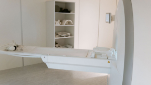
Why Quality Assurance Matters in Digital Radiography
Digital radiography delivers faster results and improved workflow, but consistent quality requires more than advanced detectors. Without structured X-ray QA, imaging centers risk inaccurate results, higher repeat rates, and unnecessary radiation exposure.
Investing in digital radiography equipment is only part of the solution—QA protocols guarantee that technology performs at its peak.
Core Elements of Medical Imaging Quality Control
Strong medical imaging quality control includes daily, weekly, and annual checks using appropriate testing equipment.
- Daily checks: Warm-up cycles, visual display checks, and verification of test leads.
- Weekly checks: Phantom imaging for diagnostic accuracy, vibration measurements on moving parts, and spectrum analysis for electronic performance.
- Monthly checks: Detector calibration with waveform generators and signal generators.
- Annual checks: Comprehensive performance evaluation using thermal imagers, Insulation Resistance Tester devices, and digital oscilloscope readings.
Together, these protocols safeguard both image quality and patient safety.
Equipment Testing and Calibration
Routine equipment testing confirms that systems remain aligned with manufacturer specifications.
- Exposure accuracy checks verify consistency across multiple shots.
- Circuit specialists may conduct power distribution inspections to confirm stable power sources.
- Waveform generators and spectrum analysis tools are used to identify performance drifts.
- Thermal imagers monitor system temperatures for signs of overheating.
Using advanced Test and Measurement Equipment ensures issues are identified before they escalate. Facilities also benefit from centralizing accessories and QA products to streamline operations.
Balancing Safety and Efficiency
While QA protects patients, it should not overburden staff. Automated testing equipment, cloud-based logs, and built-in calibration functions simplify quality control and reduce downtime.
The goal is to maintain accuracy while preserving workflow efficiency in radiology departments.
Regulatory and Accreditation Standards
Accreditation organizations require documentation of medical imaging quality control activities.
- ACR and state regulators mandate annual physicist testing with calibrated test equipment.
- Logs must show compliance with radiation safety, alignment, and exposure standards.
- Electrical equipment must pass insulation resistance and grounding checks annually.
Well-documented QA ensures facilities stay compliant and audit-ready.
Preventive Maintenance in QA Programs
Preventive maintenance supports quality assurance by combining equipment testing with servicing.
- Technicians inspect power distribution systems to reduce failure risks.
- Insulation Resistance Tester checks verify safe electrical pathways.
- Preventive vibration measurements on moving parts identify wear before breakdown.
Integrating maintenance into QA reduces downtime and extends system life. Facilities investing in modern imaging equipment should plan long-term service and testing support.
Role of Radiology Staff in QA
QA programs succeed when radiology teams participate.
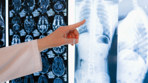
- Technologists track diagnostic image quality daily.
- Radiologists flag artifacts or subtle degradation in imaging files.
- Staff document issues and escalate them for corrective action.
Hands-on involvement helps prevent issues before they affect patients.
Leveraging Technology in QA
Modern QA uses advanced digital workflows and Test and Measurement Equipment to improve consistency.
- Digital oscilloscope tools capture performance variations.
- Spectrum analysis highlights electrical irregularities.
- Automated calibration routines ensure consistent exposures.
- Cloud platforms store QA results for secure long-term archiving.
These tools reduce reliance on manual logs and improve diagnostic reliability.
Challenges in Quality Assurance
Facilities face challenges such as limited budgets, time constraints, and differences in QA protocols across health systems.
- Smaller clinics may lack dedicated circuit specialists.
- Larger facilities may struggle with standardizing test equipment across multiple sites.
- Inconsistent calibration of power sources can affect results.
Vendor partnerships help overcome these challenges by providing guidance, training, and reliable equipment support.
Building a Culture of Quality
QA works best when it becomes part of the culture. Radiology departments must view quality assurance as essential to patient outcomes, not just a regulatory requirement.
Routine education, leadership support, and investment in testing equipment like waveform generators and thermal imagers foster this culture.
Moving Toward Continuous Improvement
QA evolves alongside technology. Facilities should update protocols as manufacturers release new recommendations, and as AI-driven systems and advanced diagnostic tools enter the market.
Continuous improvement ensures that imaging centers keep pace with innovation while maintaining patient trust.
Building Confidence in Diagnostic Imaging
So, what quality checks are required for X-ray equipment? Facilities must conduct daily readiness checks, weekly phantom imaging, monthly calibrations, and annual physicist-led evaluations with advanced test and measurement tools. This combination ensures safe, reliable, and high-quality results.
Spectrum Medical Imaging Co. has supported imaging facilities for over 40 years. From advanced X-ray QA programs to comprehensive medical imaging quality control and reliable equipment testing, our team provides unmatched expertise, service, and nationwide support. With certified technicians, same-day shipping, and trusted solutions, we help practices maintain diagnostic accuracy with confidence.