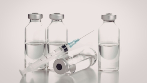
Adverse reactions during contrast media administration require immediate recognition and a structured response. Emergency response protocols must be documented, rehearsed, and accessible in every imaging suite where iodinated or gadolinium-based agents are administered. Both CT scan and magnetic resonance imaging workflows depend on contrast enhancement, so emergency readiness must account for every contrast medium routinely administered.
Standardized checklists reduce response time and eliminate ambiguity during acute events. Departments using verified emergency response frameworks demonstrate higher staff confidence and measurable improvements in patient monitoring and outcome documentation.
Key Takeaways
- Emergency response checklists include acute reaction grading, epinephrine availability verification, and extravasation management protocols
- Only 2.3% of radiology sites were rated appropriate on comprehensive safety checklists, highlighting widespread preparedness gaps
- Pre-session emergency kit verification and escalation pathway documentation must be confirmed before every contrast administration
- Severity grading determines treatment pathways, with anaphylactoid reactions requiring immediate epinephrine administration per facility protocol
- Electronic checklist systems offer stronger audit trails than manual methods, though usability challenges remain in some implementations
What Is a Contrast Media Emergency Response Checklist?
A contrast media emergency response checklist is a structured protocol defining staff actions during adverse reactions. The checklist includes acute reaction grading criteria, epinephrine availability confirmation, extravasation treatment steps, and escalation pathway activation.
Pre-administration safety components cover risk-factor screening, allergy history review, and renal function assessment. These front-end controls reduce the likelihood of severe reactions and ensure clinical teams are prepared before injection begins.
What Are the Key Components of Emergency Response in Contrast Media?
Emergency response checklists integrate multiple safety layers into a single workflow. Risk-factor screening before contrast administration identifies patients with prior hypersensitivity, renal insufficiency, or contraindicated medications.
Acute reaction grading, epinephrine availability, and extravasation management protocols form the operational core of emergency preparedness. Allergy history documentation and renal function review serve as decision checkpoints before any contrast agent is administered. Screening applies equally to iodinated agents used in CT and to gadolinium-based contrast agents administered during MRI scans, since each carries a distinct reaction profile that staff must be ready to recognize.
What Is the Clinical Importance and Patient Safety Role?
Only 2.3% of radiology sites were rated appropriate on a 49-item safety checklist in published evaluations. The remaining 97.7% of radiology sites were rated merely moderate, revealing widespread gaps in protocol completeness and staff readiness.
These findings underscore the critical role of standardized checklists in reducing preventable adverse events. Facilities without complete protocols face higher incident rates and lower staff confidence during acute situations.
The Contrast Media Emergency Response Checklist
This ten-step checklist provides a complete framework for contrast media safety and rapid protocol execution. Each step addresses a specific phase of preparation, recognition, treatment, or documentation required during contrast administration sessions.
Departments should adapt this framework to local workflows while maintaining the core sequence. All steps must be completed in order to ensure comprehensive readiness and effective radiology incident management.
Step 1: Confirm Pre-Session Emergency Kit and Medication Readiness
Emergency kits must be verified for readiness before any session involving iodinated contrast administration. Omnipaque (iohexol, GE HealthCare) is among the most frequently administered iodinated agents in CT departments.
Confirm the emergency kit is fully stocked and accessible before any contrast session begins. Verify epinephrine is available, and the kit location is known to all staff on duty.
Step 2: Confirm Escalation Pathway Is Documented and Posted
Staff must be trained to recognize and respond to reactions from any administered contrast agent. Widely used nonionic iodinated agents such as Optiray (ioversol, Guerbet) require specific reaction grading and escalation protocols.
Confirm escalation pathway documentation is current and visible in the contrast room. Confirm that all staff on duty understand their assigned roles in the escalation sequence.
Step 3: Recognize Signs and Symptoms of an Acute Contrast Reaction
Acute reactions to iodinated agents such as Isovue (iopamidol, Bracco) range from mild urticaria to severe responses. Staff must be able to recognize and categorize the full spectrum of signs on presentation.
Assess the patient immediately upon any reported or observed change in condition following contrast administration. Early recognition reduces progression risk and enables faster treatment initiation.
Step 4: Grade the Reaction Severity
Apply the acute reaction grading protocol to classify the reaction as mild, moderate, or severe. Severity grade determines the treatment pathway and escalation level activated.
Grading systems typically assess cardiovascular, respiratory, and dermatologic findings using standardized criteria. Consistent application of grading rules improves response accuracy and documentation quality.
Step 5: Stop Contrast Administration and Stabilize the Patient
Halt contrast injection immediately upon confirmed or suspected reaction. Position the patient appropriately and maintain airway, breathing, and circulation.
Patient monitoring intensifies during this phase to track symptom evolution and response to initial interventions. Stabilization procedures prevent deterioration while the treatment team assembles.
Step 6: Administer Emergency Agents Including Epinephrine
Administer epinephrine for anaphylactoid or severe reactions per facility protocol. In referral hospitals, 87.7% maintain epinephrine availability in CT rooms for immediate access during acute events.
Follow dosing and administration instructions per the graded reaction level. Epinephrine delivery routes and doses vary depending on reaction severity and patient response.
Step 7: Manage Extravasation When Present
Apply extravasation management steps when contrast leakage into the surrounding tissue is identified. Elevate the affected limb, apply a warm compress if appropriate, and monitor the site.
Document extravasation details, including volume estimate, site location, and patient response. Complete documentation supports follow-up care and departmental quality review.
Step 8: Activate Full Escalation Pathway for Severe Reactions
Escalation pathway activation for moderate-to-severe reactions includes notifying the attending physician and calling emergency response. Only 30.4% of young radiologists felt confident in the primary management of adverse reactions.
Escalation readiness must be drilled, not assumed, to ensure reliable execution during actual events. Preparing for patient transfer and coordinating with emergency teams reduces response delays.
Step 9: Apply GBCA-Specific Response Protocols When Applicable
Training programs should include GBCA-specific adverse reaction content for macrocyclic agents. Gadavist (gadobutrol, Bayer) and other macrocyclic GBCAs carry a different risk profile from iodinated agents. These gadolinium contrast agents rely on the magnetic properties of gadolinium to alter signal intensity within the magnetic field of an MRI system, so response training must address this modality directly.
Simulation drills should include scenarios covering both iodinated and gadolinium-based contrast reactions. Dotarem (gadoterate meglumine, Guerbet) is a macrocyclic GBCA requiring competency refreshers on hypersensitivity recognition.
Step 10: Complete Incident Documentation
Document reaction type, severity grade, treatment administered, patient response, and escalation actions taken. Maintain audit trails for compliance and departmental review.
Incident documentation completion is required before the session record is closed. Comprehensive records support quality improvement initiatives and regulatory compliance.
How Do Emergency Checklists Support Radiology Departments?
Emergency checklists provide standardized workflows that align staff actions with departmental policies. They reduce variability in response quality and ensure that critical steps are not omitted during high-pressure situations.
Integration with electronic health records enables real-time documentation and audit trail generation. Departments using structured checklists demonstrate higher compliance rates and improved incident outcome tracking.
How Is the Checklist Integrated With Departmental Policies and Protocols?
All hospitals screen patient risk factors before iodinated contrast administration as part of departmental policy. Fasting requirements are enforced by 98.3% of hospitals to reduce aspiration risk during adverse events.
Checklist integration ensures that individual staff actions align with facility-wide contrast media safety standards. Policy updates flow directly into checklist revisions, maintaining consistency across all imaging modalities.
What Are Staff Roles and Responsibilities in Adverse Reaction Management?
Managing allergic reactions requires clear role assignments for radiologists, technologists, and nursing staff. Reaction grading and escalation protocols should be rehearsed for each agent type in departmental use.
Staff must understand who initiates treatment, who documents findings, and who activates escalation pathways. Role clarity prevents duplication of effort and eliminates response gaps during acute events.
How Is Emergency Kit and Medication Readiness Maintained?
Emergency kits must be fully stocked and verified before any session involving iodinated contrast administration. In referral hospitals, 98.3% maintain emergency agents in CT rooms for immediate access.
Emergency kit location identification must be part of onboarding for all new imaging staff. Regular inventory audits and expiration-date checks ensure that medications remain available and effective.
What Are the Most Common Contrast Media Reactions and Their Grading?
Contrast reactions range from mild cutaneous symptoms to life-threatening cardiovascular collapse. Grading systems classify reactions by severity to guide treatment intensity and escalation decisions.
Mild reactions include localized urticaria, nausea, or brief vasovagal responses requiring minimal intervention. Severe reactions involve respiratory distress, cardiac arrhythmias, or widespread anaphylaxis requiring immediate emergency treatment.
What Signs and Symptoms Indicate an Acute Reaction?
Acute reactions to iodinated agents such as Isovue (iopamidol, Bracco) range from mild urticaria to severe responses. Staff must be able to recognize and categorize the full spectrum of signs on presentation.
Early symptoms may include flushing, pruritus, or mild dyspnea appearing within minutes of injection. Progressive symptoms such as hypotension, bronchospasm, or laryngeal edema signal severe reactions requiring escalated intervention.
How Are Severity Levels and Treatment Priorities Determined?
Acute reaction grading assessment uses standardized criteria to classify findings as mild, moderate, or severe. Cardiovascular stability, respiratory function, and dermatologic status are evaluated simultaneously.
Treatment priorities shift based on grading results, with severe reactions receiving immediate epinephrine and emergency support. Moderate reactions may require antihistamines, corticosteroids, and extended observation before discharge.
What Training and Confidence Building Are Needed for Effective Emergency Response?
Effective emergency response depends on repeated training and competency validation for all imaging staff. Knowledge gaps in reaction management create hesitation and delay during actual events.
Simulation drills replicate high-stress scenarios and build muscle memory for critical procedures. Electronic and manual checklist tools support training by providing consistent reference materials during both drills and real incidents.
How Are Knowledge Gaps in Reaction Management Addressed?
Only 58.6% of young radiologists are familiar with adverse-reaction safety protocols in international surveys. Just 30.4% of young radiologists felt confident in the primary management of adverse reactions despite awareness.
A high desire to improve knowledge was reported by 95.8% of young radiologists in the same survey. Training programs should include GBCA-specific adverse reaction content for macrocyclic agents like Gadavist.
What Is the Role of Simulation Drills and Competency Refreshers?
Simulation drills should include scenarios covering both iodinated and gadolinium-based contrast reactions. Dotarem (gadoterate meglumine, Guerbet) is a macrocyclic GBCA requiring competency refreshers on hypersensitivity recognition.
Drills improve confidence, reduce response time, and identify weaknesses in escalation pathways before actual emergencies. Competency refreshers maintain proficiency in infrequently used skills such as epinephrine administration and extravasation treatment.
What Are the Benefits of Electronic Versus Manual Checklist Tools?
Manual methods are easy to deploy but harder to aggregate incidents for quality review. Paper-based tools work well in rural or low-IT settings but offer limited version control.
Electronic methods offer audit trails and structured data capture for radiology incident management. However, usability problems can increase the burden if poorly designed systems disrupt workflow.
How Does Emergency Checklist Adoption Vary by Facility and Region?
Emergency checklist adoption depends on digital infrastructure, regulatory pressure, and facility resources. Urban hospitals with robust electronic health records deploy digital checklists more rapidly than rural counterparts.
Regional variation in preparedness standards creates disparities in patient safety outcomes. Global preparedness trends show increasing investment in standardized protocols, though implementation speed differs significantly by country.
What Are the Urban Versus Rural Digital Infrastructure Impacts?
Electronic health record adoption reaches 74% in urban settings versus 64% in rural settings. Rural facilities face lower odds of certified EHR adoption, limiting their ability to deploy electronic checklist tools.
Digital infrastructure gaps force rural hospitals to rely on manual protocols and printed workflows. This creates challenges in audit trail generation and multi-site protocol standardization.
What Compliance Challenges and Workflow Fit Issues Arise?
Workflow fit determines whether checklists are consistently used or routinely bypassed during busy periods. Poorly designed checklists that disrupt patient throughput face low adoption regardless of clinical value.
Compliance challenges include staff resistance to the documentation burden and a lack of leadership accountability. Facilities must balance thoroughness with usability to achieve sustainable checklist integration.
What Global Preparedness Trends and Examples Are Relevant?
Korean referral hospitals demonstrate high preparedness, with 98.3% maintaining emergency agents in CT rooms. International surveys reveal uneven protocol familiarity, with only 58.6% of young radiologists familiar with safety standards.
Global trends favor electronic standardization, but manual methods remain common in resource-limited settings. Countries with strong regulatory frameworks show faster adoption of comprehensive emergency response checklists.
Synthesis of Contrast Media Emergency Response Checklist Practices and Priorities
Emergency response checklists integrate pre-administration screening, acute reaction grading, and escalation protocols into unified workflows. Departments using structured checklists demonstrate higher staff confidence and better patient outcomes during adverse events.
Effective implementation requires training investment, simulation drills, and continuous competency validation. Electronic tools offer advantages in audit trails and data aggregation, though manual methods remain viable where digital infrastructure is limited.
The strongest preparedness gains come from facilities that treat checklists as living documents requiring regular updates. High-risk patient management and contrast media pricing trends continue to shape how departments allocate resources toward safety infrastructure and emergency preparedness.
Strengthen Your Contrast Media Safety Program with Spectrum Medical Imaging Co.
Spectrum Medical Imaging Co. supports radiology departments with reliable imaging equipment and comprehensive lifecycle support. Our 30+ years of experience in medical imaging enable facilities to maintain operational readiness and patient safety. As a prime dealer for Guerbet, Bayer, Bracco, GE HealthCare, and Fresenius-Kabi, we deliver guaranteed lowest pricing on Omnipaque, Isovue, Optiray, Visipaque, Gadavist, Dotarem, and the other contrast agents your emergency-ready protocols depend on, backed by 24–48 hour shipping and 24/7 technical support.
Strengthen your department’s emergency preparedness and ensure your imaging equipment meets the highest safety standards. Contact our team to discuss how we can support your contrast media safety program and equipment needs.