Key Takeaways
- AI in radiology improves diagnostic accuracy by flagging subtle findings such as fractures, lung nodules, and tumors that support, not replace, the radiologist’s read.
- AI-powered X-ray systems speed image analysis and reporting, cut turnaround times, and help teams triage urgent cases faster.
- AI standardizes image interpretation, reduces inter-observer variability, and lowers costs by limiting repeat imaging and misdiagnosis.
- Human oversight, data privacy, and clear regulatory frameworks keep AI safe; radiologists stay essential for clinical judgment and patient care.
- AI-ready imaging depends on modern hardware; Spectrum Medical Imaging Co. supplies the digital radiography panels, X-ray systems, and PACS these workflows run on. Call 800-859-6162.
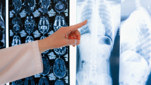
AI in radiology is changing how clinicians diagnose, interpret, and manage patient care. Artificial intelligence is moving quickly through health care, and radiology sits at the front of that change. In X-ray imaging, AI adds speed, precision, and consistency to the work radiologists already do. For facilities that want to strengthen their X-ray service, AI brings a practical mix of accuracy and efficiency. The technology runs on modern imaging hardware, which is where Spectrum Medical Imaging Co. supports facilities with digital radiography equipment and 30+ years of experience in medical imaging.
In the sections below, we walk through the main benefits of AI in X-ray imaging, from diagnostic accuracy to workflow and cost, how the tools work, the hardware they depend on, and the oversight that keeps them safe and useful.
AI in Radiology: Enhanced Diagnostic Accuracy
One of the clearest advantages of AI in radiology is better diagnostic accuracy. AI algorithms analyze X-ray images with high precision and can flag subtle abnormalities that a busy human eye may miss. The software does not make the diagnosis. It points the radiologist to areas worth a closer look.
By helping detect conditions such as fractures, tumors, and lung diseases, AI reduces diagnostic errors and raises confidence in clinical decisions. In diagnostic radiology, a second set of pattern-trained eyes on every image supports more reliable reads. The radiologist still interprets the medical images, but with an assist that catches outliers. The result is more consistent diagnostic quality and better patient outcomes.
AI in Radiology and Faster Image Analysis and Reporting
Time drives medical diagnostics. AI-powered X-ray systems can process and interpret images in seconds, which cuts turnaround times sharply. For a facility running a high-volume X-ray service, that speed reshapes the day.
The workflow gains are concrete:
- Quicker diagnoses
- Reduced patient wait times
- Improved workflow efficiency across imaging procedures
AI can also rank a worklist by urgency. With patient triage built into the queue, radiologists address critical cases first instead of working strictly in order. That prioritization matters most in emergency and trauma settings, where minutes count.
Improved Workflow Efficiency in Imaging Departments
AI streamlines many repetitive tasks across radiology departments, including image sorting, labeling, and preliminary analysis. Handling that routine load frees radiologists to focus on complex cases and patient-facing care.
The benefits build on each other:
- Reduced administrative burden
- Better resource allocation across clinical workflows
- Higher productivity within imaging departments
AI also connects to the systems a department already runs. When AI tools feed results into the PACS and link with electronic health records, findings move with the patient instead of sitting in a silo. Spectrum supplies PACS, HIS, and RIS systems that support this kind of electronic image exchange, so a facility can adopt AI without rebuilding its workflow.
Early Detection of Disease With AI X-Ray Imaging
AI is strong at recognizing patterns in imaging data, which makes it useful for early disease detection. In X-ray imaging, AI can help surface early signs of conditions like lung cancer, tuberculosis, and cardiovascular abnormalities. It can also assist with lung nodules and tumour detection that are easy to overlook at a glance.
Early detection improves treatment outcomes and lowers downstream costs. Pattern recognition extends to other modalities too, where AI helps screen for breast cancer, brain tumors, and even harder targets like pancreatic cancer. As AI identifies imaging biomarkers across studies, it feeds the broader move toward precision medicine, where care is matched to the individual patient.
Standardization, Consistency, and Cost Savings
Variability in image interpretation is a real challenge in radiology. AI introduces standardization by applying consistent criteria across every X-ray analysis. That consistency produces uniform diagnostic quality, less inter-observer variability, and more predictable patient outcomes. For any organization providing an X-ray service, reliable reads strengthen trust.
The financial case follows the quality case. AI requires an upfront investment, but it pays back over time by reducing errors and unnecessary repeat imaging. Fewer misdiagnoses lower liability risk, optimized workflows reduce operating expense, and higher throughput supports revenue. Avoiding repeat exposures also reduces wear on equipment, and Spectrum’s imaging equipment repair and maintenance helps keep that hardware dependable so AI tools have clean images to work with.
How AI X-Ray Tools Work
Most AI in radiology is built on machine learning, and specifically deep learning. Convolutional neural networks, a type of deep learning model suited to images, learn from large sets of labeled studies to recognize patterns tied to disease. This is the engine behind computer-aided diagnosis, often called CAD systems, which mark suspicious regions for review.
The toolkit has grown well beyond pattern flags. Image segmentation outlines organs and lesions so measurements are consistent. Natural language processing reads and structures radiology reports, turning free text into searchable data. Predictive analytics estimates risk from imaging and clinical inputs. Newer generative AI and multimodal AI models combine images with text and other data, and AI-supported image reporting drafts preliminary findings for the radiologist to confirm. Together, these AI-driven technologies move toward true AI-aided X-ray interpretation, where machine intelligence and human expertise work side by side.
The Imaging Hardware Behind AI
AI is only as good as the images it receives, so the hardware foundation matters. Clean image acquisition starts with the equipment in the room. An X-ray tube produces the beam, an automatic exposure device controls the dose, an anti-scatter grid sharpens contrast, and the image receptor captures the result.
The detector is central. Modern rooms use a digital flat-panel detector rather than older computed radiography or an analog X-ray image intensifier, because a clean digital signal gives AI better data to read. Emerging photon-counting detectors and dual-energy imaging add even more information per exposure, and spectral shaping filters help manage dose and image quality. Advanced workflows lean on this hardware too, from electronic fluoroscopy systems and image stitching for long-length studies to 3D imaging and digital mobile radiography equipment used at the bedside and in hybrid operating rooms.
This is where an equipment partner fits the AI conversation. Spectrum is an X-ray equipment supplier and stocks modern digital radiography panels across Southern California. Strong image processing and a quality detector give any AI tool the diagnostic-grade input it needs, so upgrading the hardware is often the first practical step toward AI-ready imaging.
Human Oversight, Standards, and the Radiologist’s Role
A common worry is that AI will replace radiologists. In practice, AI is a support tool that extends human expertise. Radiologists remain essential for clinical judgment, complex case evaluation, and patient communication. Radiologic technologists and the medical radiologist stay at the center of care, with AI handling the repetitive load.
Responsible use depends on guardrails. Human oversight keeps a clinician accountable for every result, since an AI model can behave like a black box whose reasoning is hard to inspect. Clear regulatory frameworks, transparency tools such as model cards, and strong data privacy protect patients as adoption grows. AI is most useful as clinical decision support, informing the radiologist rather than deciding for them. Sound radiology training and cross-modality education help teams use these tools well, which keeps the focus where it belongs: on accurate reads and good patient care.
Related Reading
- Digital radiography panels in Los Angeles for DR detector upgrades and replacements.
- Medical imaging products and supplies across contrast media, detectors, and equipment.
- About Spectrum Medical Imaging Co. and its 30+ years in medical imaging supply.
What to Weigh Before Adopting AI in X-Ray Imaging
Adding AI is a project, not a switch. A few practical factors decide whether it pays off. Image quality comes first, since an AI tool trained on clean studies needs clean studies to perform. A facility running aging detectors or computed radiography often sees better returns by upgrading the hardware before layering on software.
Integration is the next question. AI results are only useful if they reach the radiologist inside the existing workflow, so the tool should connect with the PACS and electronic health records the team already uses. Regulatory clearance matters too: confirm that any AI product carries the appropriate clearance for clinical use, and plan for the validation and human oversight that keep it accountable. Finally, budget for staff training, since radiologic technologists and radiologists need time to learn how a new tool fits their reads. Treating AI as part of a wider imaging upgrade, rather than a standalone add-on, gives the technology the foundation it needs.
Build the Imaging Foundation Your AI Tools Need
AI in radiology delivers its value only when it runs on dependable, diagnostic-grade imaging. Spectrum Medical Imaging Co. supplies the digital radiography panels, X-ray systems, and PACS that AI-ready workflows depend on, backed by 30+ years in medical imaging and a focus on Southern California and the Los Angeles metro. We do not sell hype. We help your facility put the right hardware and image-management foundation in place, so any AI tools you add have clean data and a stable workflow to build on.
Talk with a specialist about modernizing your X-ray and digital radiography equipment. Call 800-859-6162 or request a quote to get started. Explore the full medical imaging line at spectrumxray.com.
Frequently Asked Questions
How does AI improve X-ray diagnostic accuracy?
AI algorithms are trained on large sets of labeled images, so they recognize patterns tied to conditions like fractures, lung nodules, and tumors. During a read, the software flags subtle findings that a busy radiologist might overlook and marks regions for closer review. It does not make the diagnosis; the radiologist still interprets the image. This second-look support reduces diagnostic errors and improves confidence in the final read.
Will AI replace radiologists?
No. AI is a support tool that augments human expertise rather than replacing it. Radiologists remain essential for clinical judgment, complex case evaluation, and patient communication, and they stay accountable for every result. AI handles repetitive tasks and flags findings, while the radiologist makes the call. The most effective model pairs machine intelligence with human oversight.
What kinds of AI are used in radiology?
Most radiology AI uses machine learning, especially deep learning models called convolutional neural networks that are well suited to images. These power computer-aided diagnosis, image segmentation, and predictive analytics. Natural language processing structures radiology reports, while newer generative and multimodal AI combine images with other data. All of it is meant to assist the radiologist as clinical decision support.
Does AI X-ray imaging require new equipment?
AI software needs clean, diagnostic-grade images to work well, so the imaging hardware matters. A modern digital flat-panel detector produces a clean digital signal that gives AI better data than older computed radiography or analog image intensifiers. Many facilities find that upgrading the detector and PACS is the practical first step toward AI-ready imaging. Spectrum Medical Imaging Co. supplies the digital radiography panels, X-ray systems, and PACS that support these workflows.
Is AI in radiology safe and regulated?
AI in radiology is designed to operate under human oversight, with a clinician accountable for every result. Regulatory frameworks, transparency tools such as model cards, and strong data privacy protections govern how these tools are used. Because an AI model can act like a black box, clinical validation and ongoing monitoring matter. Used as clinical decision support, AI informs the radiologist rather than deciding for the patient.