Key Takeaways
- Well-designed Guerbet Optistar CT protocols directly link image quality, patient safety, and injector longevity by aligning flow, volume, and timing with scanner capabilities.
- OptiVantage and OptiOne support multi-phase, automated protocols that reduce manual steps, contrast waste, and re-scans while maintaining high diagnostic success rates.
- OptiBolus® decelerating flow profiles can cut contrast load by up to 40% while sustaining uniform vascular enhancement across diverse CT indications.
- Data from injector logs, contrast usage, and repeat-scan rates enable continuous refinement of protocols to standardize enhancement and reduce variability.
- Indication-specific and patient-tailored protocols—especially for oncology, emergency, pediatric, and frail patients—optimize clinical outcomes while limiting mechanical stress on injectors.
Advanced CT contrast protocols sit at the intersection of image quality, patient safety, and scanner efficiency. When timing, flow rate, and volume are optimized, radiology teams capture diagnostic detail on the first attempt while avoiding repeat scans, excess contrast use, and unnecessary injector wear. Guerbet’s Optistar family, including OptiVantage and OptiOne, is engineered to translate protocol precision into consistent, reproducible enhancement across diverse CT indications. This article explains how protocol design shapes enhancement, how Optistar hardware and software support multi-phase workflows, and how data-driven refinement can extend injector performance while controlling costs and improving patient experience in busy CT departments.
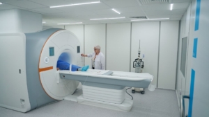
What Is the Guerbet Optistar CT Injector and Why Does Protocol Design Matter?
Guerbet Optistar CT protocols directly impact diagnostic quality and departmental throughput. The Optistar family—OptiVantage and OptiOne—represents Guerbet’s approach to advanced CT imaging optimization, where hardware capabilities meet protocol precision. Understanding how these injectors integrate into modern workflows reveals why contrast injection timing and flow parameters drive both image quality and operational efficiency.
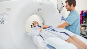
How Does the Optistar Injector Operate Within Modern CT Scan Workflows?
OptiVantage is a dual-head CT contrast delivery injector built for complex injection protocols that match today’s high-speed CT scanners. OptiOne offers single-head functionality compatible with prefilled syringes and consumable systems. Both platforms accept prefilled syringes or standard contrast media vials, enabling tailored CT exams across patient populations. The systems improve CT workflow efficiency through streamlined interfaces, automated operations, and simplified handling protocols that reduce technologist workload while maintaining injection precision.
What Are “Advanced” CT Contrast Protocols in Practical, Day-to-Day Terms?
Advanced protocols mean multi-phase automated injection sequences that deliver contrast at precise intervals without manual intervention. OptiOne extends this capability beyond standard CT suites into radiotherapy, trauma scanners, mammography, and PET CT environments. Advanced Guerbet Optistar CT protocols automate what previously required manual coordination—phase timing, volume changes, and saline chasers—allowing technologists to focus on patient care rather than injection mechanics.
How Do Protocol Choices Influence Both Image Quality and Injector Workload?
Protocol optimization directly determines diagnostic success. A recent clinical study demonstrated 100% successful diagnostic imaging with OptiVantage in multi-patient mode (95% CI: 95.39%, 100.00%) across common indications, including breast, colon, and lung cancer imaging. Well-designed protocols reduce repeat scans, minimize contrast waste, and decrease injector wear from unnecessary high-pressure injections. Poor protocols force re-scans, waste contrast media, and stress hardware—making protocol design a key radiology equipment performance tip that affects both clinical outcomes and equipment longevity.
How Do CT Contrast Injection Parameters Shape Image Quality and Diagnostic Confidence?
Contrast injection timing and flow parameters determine whether CT scans achieve diagnostic quality on the first attempt. Volume, flow rate, and injection duration must align with patient physiology and scanner capabilities to capture optimal vascular and organ enhancement. Advanced CT imaging optimization requires understanding how these variables interact—and how Guerbet Optistar CT protocols use this knowledge to standardize enhancement across diverse patient populations.
How Do Volume, Flow Rate, and Injection Duration Affect Vascular and Organ Enhancement?
Clinical data show mean injected volumes of 119.5 ± 14.4 mL with injection rates between 2.8-4.5 mL/s (mean: 3.6 ± 0.3 mL/s) achieve consistent diagnostic results. OptiBolus® technology employs exponentially decelerating flow rate injection, extending the period of uniform vascular enhancement compared to constant-rate protocols. This approach maintains image quality while reducing total contrast load—uniform enhancement matters more than peak enhancement for most diagnostic applications. The decelerating flow profile ensures target anatomy remains enhanced throughout the scan acquisition window.
How Do Patient Factors Such as Weight, Cardiac Output, and Renal Function Alter Protocol Design?
Real-world imaging populations are diverse: 59% women, mean age 63.6 ± 12.7 years (range 18-83), with 55% over age 65. OptiBolus reduces overall contrast load by up to 40%, though specific automated patient-size-based dose modulation is not explicitly detailed in the current Guerbet documentation. This general contrast reduction represents a significant step toward patient-specific dose optimization—lower volumes benefit patients with renal concerns while maintaining diagnostic confidence. Effective Guerbet Optistar CT protocols account for patient variability even without fully automated weight-based adjustments.
How Do Scanner Settings (kVp, Rotation Time, Detector Configuration) Interact with Contrast Protocols?
Guerbet CT injectors are designed to keep pace with increasing CT scanner speeds—modern scanners acquire data faster than older systems, demanding precise contrast injection timing and flow synchronization. Scanner settings like kVp (which affects contrast visibility), rotation time (which determines acquisition speed), and detector configuration (which impacts temporal resolution) all influence when contrast must arrive in target vessels. Optimal enhancement during specific phases of organ or vessel opacification requires protocols that account for both injector capabilities and scanner parameters—a key radiology equipment performance tip for maximizing diagnostic yield.
How Does Guerbet Optistar Support Flexible, Protocol-Driven CT Contrast Delivery?
Protocol flexibility separates basic injectors from systems that adapt to diverse clinical demands. Guerbet Optistar CT protocols leverage dual-head design, programmable multi-phase sequences, and intuitive interfaces to deliver consistent results across complex imaging scenarios. This flexibility directly improves CT workflow efficiency—technologists spend less time managing injections and more time managing patients.
How Can Optistar’s Dual-Head Design Be Used for Saline Chasers and Multi-Phase Studies?
OptiVantage’s dual-head design enables bi-liquid studies where contrast and saline flow independently without manual switching. Saline chasers push residual contrast from tubing into the patient, maximizing iodine delivery efficiency and reducing waste. The ability to program multiple phase protocols ensures contrast delivery synchronizes with CT acquisition phases—arterial, venous, and delayed phases execute automatically at predetermined intervals. This automation eliminates timing errors that compromise image quality in CT angiography and multi-phase abdominal studies.
How Do Pre-Set and Custom Protocols Simplify Complex CT Angiography and Multiphase Exams?
Clinical validation from Chelsea Westminster Hospital NHS Foundation Trust and Liverpool Heart & Chest Hospital NHS Foundation Trust demonstrates real-world OptiBolus effectiveness across diverse patient populations. Pre-set protocols encode institutional best practices for common indications—technologists select the appropriate exam type and the system applies proven contrast injection timing and flow parameters. Custom protocols allow adaptation for unusual patient factors or specialized research applications. This balance between standardization and flexibility represents advanced CT imaging optimization—consistent quality without sacrificing clinical judgment.
How Do Optistar Interface Features Help Technologists Apply Protocols Consistently?
Interface design directly impacts CT workflow efficiency. Patient preparation time averages 6-10 seconds for 68% of cases and 16-20 seconds for 30% of patients in clinical studies. Dayset changes—swapping disposable components between patients—complete within one minute. Streamlined workflows reduce technologist stress and minimize opportunities for protocol errors. Clear interface prompts, logical menu structures, and visual confirmations ensure protocols execute as intended—a critical radiology equipment performance tip for departments seeking to reduce variability and improve throughput without compromising safety.
How Can Optimized Contrast Protocols Improve Imaging Quality and Extend the Performance of Guerbet Optistar CT Injectors?
Protocol optimization delivers dual benefits: better diagnostic images and longer equipment life. Well-designed Guerbet Optistar CT protocols reduce mechanical stress on injector components while improving vascular enhancement consistency. This connection between clinical quality and equipment longevity makes protocol refinement a priority for departments seeking both diagnostic excellence and cost control.
How Can Weight-Based and Iodine-Load–Based Protocols Standardize Enhancement Across Patients?
OptiBolus® technology reduces contrast load by up to 40% without compromising diagnostic confidence—a significant achievement in advanced CT imaging optimization. Bolus shaping software optimizes contrast usage through controlled flow profiles that provide extended periods of uniform vascular enhancement. This approach standardizes enhancement across patients of different sizes and physiologies without requiring explicit weight-based calculations. Reducing contrast volume while maintaining diagnostic quality represents the ideal outcome: lower costs, reduced patient exposure, and consistent image quality.
How Does Matching Bolus Timing to Specific CT Indications Reduce the Need for Repeat Scans?
Clinical studies demonstrate 100% successful diagnostic imaging with OptiVantage in multi-patient mode—zero repeat scans needed. Precise contrast injection timing and flow matched to specific clinical indications (oncology staging, vascular imaging, organ-specific protocols) ensures target anatomy enhancement at the exact moment the scanner acquires data. Mistimed injections force repeat scans, doubling contrast exposure and radiation dose while wasting time and resources. Optimized timing eliminates this waste—a critical radiology equipment performance tip for quality improvement initiatives.
How Can Smoother Flow Profiles and Appropriate Pressure Limits Reduce Stress on Patient Veins and Injector Hardware?
Exponentially decelerating flow rates maintain consistent contrast levels in target anatomy while reducing peak injection pressures. High constant-flow injections stress patient veins (increasing extravasation risk) and injector mechanical components (accelerating wear). Optimized injection profiles reduce these risks—gentler on patients and equipment simultaneously. Lower peak pressures extend injector component life while improving patient safety, demonstrating how CT workflow efficiency and equipment longevity align when protocols are properly designed.
How Does Minimizing Contrast Waste and Unnecessary Injections Support Injector Longevity and Reliability?
OptiVantage Multi-use configuration saves approximately 35 minutes per shift when performing 20 injected bi-liquid studies compared to single-patient use—a 64% faster workflow. This efficiency reduces total injector cycles per day, decreasing mechanical wear. Industry-wide contrast waste reduction potential reaches 50-59% with optimized iodinated contrast media inventory management according to ACR guidelines. Fewer unnecessary injections mean fewer mechanical cycles, less disposable waste, and extended injector service intervals. Protocol optimization thus becomes a maintenance strategy—reducing operational costs while extending capital equipment life through intelligent use rather than reactive repairs.
How Should CT Teams Structure Advanced Contrast Protocols for Different Clinical Indications?
Clinical indications determine protocol requirements—one-size-fits-all approaches compromise either image quality or efficiency. Guerbet Optistar CT protocols must adapt to routine body imaging, complex vascular studies, oncology surveillance, and emergency trauma scenarios. Structuring protocols by indication ensures appropriate contrast injection timing and flow for each clinical question while maintaining departmental workflow standards.
How Should Protocols Differ for Routine Body CT, CT Angiography, and Perfusion Studies?
OptiBolus technology provides flexibility for different enhancement needs across imaging types. Routine body CT requires moderate flow rates and single-phase acquisition—straightforward protocols optimized for solid organ visualization. CT angiography demands higher flow rates and precise bolus timing to capture peak arterial enhancement in target vessels. Perfusion studies require specialized low-volume, prolonged-injection protocols that maintain steady contrast levels during dynamic acquisition. Complex injection protocols for specialized studies leverage OptiVantage’s programmable multi-phase capabilities, delivering different volumes at different rates during a single exam—advanced CT imaging optimization for sophisticated clinical questions.
How Can Protocols Be Tailored for Oncology Follow-Up Versus Acute Emergency Imaging?
OptiOne’s design for multiple imaging environments, including a trauma scanner, demonstrates protocol versatility requirements. Oncology follow-up protocols prioritize consistency—comparing current scans to prior studies requires standardized enhancement to detect subtle interval changes. Acute emergency imaging prioritizes speed—abbreviated protocols with rapid bolus delivery enable faster diagnosis in trauma or stroke evaluation. Multi-phase protocols allow customized approaches to different clinical scenarios: extended delayed phases for genitourinary imaging, rapid arterial-only phases for pulmonary embolism, or multi-phasic liver protocols for hepatic lesion characterization. This adaptability represents a key radiology equipment performance tip—matching protocol complexity to clinical urgency.
How Should Pediatric and Frail Patient Protocols Balance Dose, Safety, and Image Quality?
ACR guidelines emphasize the importance of patient size in radiation dosimetry and contrast administration—pediatric and frail patients require dose reduction without sacrificing diagnostic adequacy. Guerbet Optistar’s ability to support tailored CT exams contributes to personalized contrast delivery strategies. Lower flow rates reduce extravasation risk in small or fragile veins. Reduced contrast volumes minimize renal exposure in elderly patients with borderline kidney function. Weight-based protocols ensure appropriate iodine delivery for smaller patients—CT workflow efficiency must accommodate special populations without creating parallel workflow systems that confuse technologists or slow operations.
How Can Data from Optistar Logs and CT Systems Guide Protocol Optimization Over Time?
Data-driven protocol refinement transforms one-time improvements into continuous performance gains. Optistar injector logs and CT system data reveal patterns invisible to individual technologists—repeat scan rates, contrast utilization trends, and workflow bottlenecks that respond to systematic intervention. Monitoring key performance indicators enables objective assessment of Guerbet Optistar CT protocols and guides evidence-based adjustments that improve both clinical outcomes and CT workflow efficiency.
Which KPIs (HU Targets, Repeat-Scan Rates, Contrast Usage) Should Teams Monitor Routinely?
Adverse event rates represent the primary safety metric—a 2025 OptiVantage multi-patient mode study reported 0% adverse events, including extravasation, air embolism, and sepsis (95% CI: 0.00%-3.62%). User satisfaction exceeding 96% in recent clinical studies indicates protocol usability and technologist confidence. Workflow efficiency metrics available from system data include exam completion times, protocol execution errors, and contrast volume per exam type. Repeat-scan rates signal mistimed injections or inadequate enhancement. Monitoring Hounsfield unit (HU) targets in major vessels ensures consistent enhancement across patients—falling below institutional thresholds indicates protocol adjustment needs. These KPIs transform subjective impressions into quantifiable radiology equipment performance tips.
How Can Injection and Dose Data Be Used to Refine Protocols and Reduce Variability?
Clinical data support that bolus shaping software reduces contrast without reducing diagnostic confidence—all injections in published studies achieved diagnostic quality. Analyzing injection data reveals whether certain patient populations consistently require protocol modifications or whether protocols perform uniformly. Dose data identifies opportunities for contrast reduction in specific indications where current protocols overdeliver iodine. Tracking contrast injection timing and flow parameters against diagnostic success rates enables evidence-based protocol refinement—tightening timing windows, adjusting flow rates, or modifying volumes based on actual outcomes rather than theoretical assumptions. This represents advanced CT imaging optimization through continuous measurement and adjustment.
How Can Quality Improvement Cycles Turn Protocol Changes into Lasting Performance Gains?
Continuous updates and ongoing clinical studies underscore Guerbet’s commitment to enhancing efficiency, safety, and image quality. OptiVantage Multi-use documentation updated September 2025 and OptiOne updated February 2025 reflect evolving best practices. Quality improvement cycles follow a standard pattern: measure baseline performance, implement protocol changes, measure outcomes, standardize successful modifications, and repeat. Lasting gains require documentation of protocol versions, training on changes, and periodic audits to prevent protocol drift. Regular review meetings where radiologists and technologists discuss diagnostic quality, workflow issues, and patient outcomes keep protocols aligned with clinical needs. This systematic approach transforms individual improvements into institutional knowledge that survives staff turnover and equipment upgrades.
Driving CT Performance Forward With Smarter Protocols
Advanced Guerbet Optistar protocols show that better images, safer injections, and longer injector life all come from the same place: deliberate, data-driven design. By standardizing iodine load, matching bolus timing to indication, and reducing wasteful injections, CT teams can cut repeat scans while protecting patients and equipment. At Spectrum Medical Imaging Co., we help you review current protocols, translate Optistar capabilities into practical workflows, and track KPIs that prove the value of change. Partner with us to turn your CT injector from a basic pump into a strategic performance and safety asset.