Key Takeaways:
- Amorphous silicon detectors typically last 7–10 years, degrading faster than generators and tubes, making panel replacement a distinct planning consideration.
- Image quality degradation—noise, artifacts, and resolution loss that cannot be corrected through calibration—indicates sensor fatigue requiring replacement.
- The European Society of Radiology recommends replacing equipment older than 10 years, as it is no longer considered state-of-the-art.
- Modern detectors reduce radiation dose while improving image clarity through increased sensitivity and advanced signal processing.
- When annual maintenance costs exceed 15–20% of replacement cost, replacing the aging panel delivers better clinical and economic value.
Aging DR panels compromise diagnostic accuracy gradually. The decline happens slowly—noise increases, artifacts appear, pixel defects multiply. By the time problems become obvious, diagnostic quality has already suffered. Radiologists may not realize their interpretations have become less reliable. Technologists adapt to worsening equipment without recognizing the cumulative impact. Understanding how panel aging affects accuracy helps facilities recognize problems early and take action before patient care suffers.
This guide explains the connection between detector aging and diagnostic performance, and shows how timely replacement restores the accuracy your radiologists need.
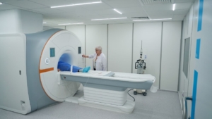
What Does Aging X-Ray Equipment Mean in the Context of DR Panel Performance?
Aging affects DR panels differently from other X-ray system components. Detectors degrade faster than generators and tubes due to the sensitive nature of their electronic and optical components. Understanding what ages and why helps facilities anticipate problems before they affect diagnostic quality and plan replacement accordingly.
How does detector aging differ from general X-ray system aging?
Detectors wear faster than other system components. The core of a DR panel is its detector technology, which falls into two main categories: indirect conversion using amorphous silicon with CsI scintillator, and direct conversion using amorphous selenium. Indirect conversion detectors using amorphous silicon typically last 7–10 years, while amorphous selenium detectors may have shorter lifespans. Generators and tubes often outlast panels by years. This difference makes panel replacement a distinct planning consideration from full system replacement.
Which components inside a DR panel naturally degrade over time?
Multiple components degrade simultaneously inside aging panels. Amorphous silicon photodiode arrays and scintillator materials experience cumulative exposure damage with every exam. Electronics, circuits, and wireless communication components deteriorate with age and continuous use. Pixel elements fail progressively, expanding from individual defects to larger non-responsive zones over time. No single component fails—the entire detector degrades as a system.
How does declining detector performance influence overall diagnostic accuracy?
Detector performance directly determines diagnostic capability. DR systems have finite lifespans, and understanding when to replace components is crucial for maintaining diagnostic accuracy, ensuring patient safety, and managing long-term operational costs. Declining detector performance compromises diagnostic quality and operational efficiency simultaneously. Image quality suffers first. Workflow problems follow. Eventually, diagnostic accuracy becomes unreliable.
How Can You Identify When a DR Panel Is Showing Early Signs of Aging?
Early detection enables planned replacement. Waiting for obvious failure delays action until diagnostic accuracy has already suffered. Recognizing subtle signs allows a proactive response.
What image-quality changes—noise, banding, artifacts—signal detector deterioration?
Image quality changes signal underlying detector problems. Image quality degradation manifests as increased noise, artifacts, inconsistent contrast, or resolution loss that cannot be corrected through routine calibration. When a panel consistently produces images with high noise, low contrast, or artifacts that interfere with diagnosis, it indicates sensor fatigue or damage. Calibration may temporarily mask symptoms but cannot reverse underlying deterioration. Persistent image quality problems require replacement consideration.
How do pixel defects evolve from individual failures to disruptive clusters?
Pixel failures multiply predictably over time. Dead pixels or non-responsive areas on the panel can obscure vital diagnostic information critical for accurate interpretation. Individual pixel failures multiply into larger non-responsive zones as the detector ages, expanding from isolated defects to disruptive clusters. Track pixel defect maps over time. Accelerating failure rates indicate approaching end-of-life even when current defect counts seem acceptable.
How do dose drift and inconsistent exposure response appear in clinical images?
Do drift signals decline detector efficiency? Increased radiation dose required to achieve acceptable image quality points to declining detector efficiency and compromises patient safety. Clinical images that are consistently blurry, grainy, or distorted indicate the detector is nearing the end of its life. Technologists compensate with higher doses, violating ALARA principles. When consistent technique produces inconsistent results, the detector—not the technique—is the problem.
What Operational Symptoms Suggest That Panel Aging Is Affecting Diagnostic Output?
Operational problems accompany image quality decline as panels age. Workflow disruptions increase costs and reduce efficiency measurably. These symptoms often appear before image quality problems become obvious to clinical users, making them valuable early warning indicators.
How do repeated exams and rising rejection rates signal reduced diagnostic reliability?
Rising repeat rates directly indicate declining diagnostic reliability. Image quality problems force technologists to repeat exams, increasing patient dose and reducing workflow efficiency across busy shifts. Track repeat rates by room and detector systematically. Compare current rates against historical baselines regularly. Rising rejection rates signal declining panel performance, affecting diagnostic output reliability before radiologists formally complain. A 5% increase in repeat rates may seem minor but compounds into significant dose and efficiency impacts over thousands of annual exams.
How do intermittent wireless connectivity issues indicate aging internal electronics?
Communication failures indicate hardware degradation beyond the detector itself. Software incompatibilities with modern EHR systems or PACS can create workflow bottlenecks and security vulnerabilities that disrupt operations significantly. Intermittent disconnects and communication failures indicate degrading internal electronics and circuitry within the panel. Wireless panels experience increasing connection instability as components age and deteriorate. These operational symptoms often precede obvious image quality decline by months, providing an early warning opportunity.
How does increased downtime or frequent resets reflect end-of-life behavior?
Increasing downtime signals systemic failure approaching. Increased downtime due to frequent equipment errors, system freezes, or hardware malfunctions is a sign that a panel has reached the end of its life. As equipment ages, the risk of breakdown and failure rises, leading to more frequent service calls and operational disruptions that affect patient scheduling. When resets become routine rather than exceptional, the panel is approaching end-of-life regardless of current image quality. Track reset frequency over time to identify accelerating trends.
How Do QC Tests and Physics Evaluations Reveal Declines in DR Panel Accuracy?
QC testing provides objective performance data over time. Clinical observation alone misses subtle degradation. Systematic testing reveals problems before they affect patient care.
Which QC metrics (uniformity, SNR, DQE) directly correlate with diagnostic accuracy?
Specific QC metrics predict diagnostic capability directly. QC tests track detector uniformity, signal-to-noise ratio, and exposure response over time through standardized measurements. These metrics directly correlate with the panel’s ability to produce diagnostically accurate images. Declining uniformity creates inconsistent image quality across the detector surface. Falling SNR reduces subtle detail visibility. DQE degradation means more dose for equivalent image quality.
How do physicists detect subtle detector aging before clinical users notice?
Physics testing reveals problems invisible to clinical observation. Physics evaluations identify performance issues not apparent in daily clinical use through specialized testing protocols designed to stress detector capabilities. Physicist testing detects subtle degradation before it affects diagnostic quality or triggers compliance failures. Annual physics surveys provide an independent assessment. Physicists recognize degradation patterns from experience across many detector installations.
How can long-term QC trend analysis determine whether a detector is still safe to use?
Trending data reveals a gradual decline that snapshot testing misses. When a panel fails to meet regulatory or diagnostic standards set by the FDA or state health departments, replacement becomes a matter of compliance as well as clinical necessity. Trending QC data reveals a gradual performance decline that clinical observation alone might miss entirely. Compile trends over months and years. Compare against manufacturer specifications and regulatory thresholds systematically.
Why Does Detector Aging Directly Reduce Diagnostic Confidence and Precision?
Diagnostic confidence depends on image quality. Aging detectors produce images that undermine radiologists’ performance. Understanding this connection clarifies why replacement matters clinically.
How does increased noise obscure fine anatomical detail and subtle pathology?
Noise directly reduces diagnostic visibility. Increased noise obscures fine anatomical detail and subtle pathologies that radiologists need to identify for accurate diagnosis. Noise degrades the diagnostic value of images, reducing radiologists’ confidence in findings. Subtle findings disappear into noisy backgrounds. Radiologists hedge their interpretations when image quality is uncertain. Missed findings and delayed diagnoses result from inadequate image quality.
How do artifacts distort anatomy and mislead radiologist interpretation?
Artifacts create diagnostic confusion. Artifacts that interfere with diagnosis indicate sensor fatigue or damage requiring immediate attention. Artifacts distort anatomical structures and can mislead radiologists’ interpretation, potentially causing diagnostic errors. Artifacts may mimic pathology or obscure real findings. Radiologists must distinguish real anatomy from artifact contamination. This additional cognitive burden reduces interpretation accuracy and efficiency.
How does an inconsistent detector response compromise the reproducibility of findings?
Inconsistent response prevents reliable comparison. Inconsistent contrast and exposure response prevent reproducible imaging results across serial exams. Technologists cannot trust that the proper technique will produce acceptable images consistently. Follow-up exams cannot be compared reliably when detector response varies unpredictably. Diagnostic confidence requires reproducible imaging. Aging detectors undermine this fundamental requirement.
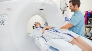
When Does DR Panel Aging Begin to Compromise Patient Outcomes?
Patient outcomes depend on diagnostic accuracy. Panel aging affects patients through multiple pathways. Understanding these connections justifies replacement investment.
How does reduced image clarity increase the chance of missed or delayed diagnoses?
Reduced clarity directly increases diagnostic risk. Resolution loss and image degradation increase the risk of missed or delayed diagnoses with real patient consequences. This is particularly critical in orthopedics and mammography, where precise imaging is essential for accurate diagnosis. Subtle fractures disappear into noisy images. Early cancers become invisible when resolution degrades. Patient outcomes suffer when diagnostic accuracy fails.
How does panel degradation lead to unnecessary radiation exposure from repeat exams?
Degraded panels increase patient dose unnecessarily. Declining detector efficiency requires increased radiation dose to achieve acceptable image quality for each exam. Repeat exams due to poor image quality compound patient radiation exposure beyond necessary levels. Both factors violate ALARA principles. Patients receive more radiation than necessary when detectors degrade. This preventable exposure accumulates over time.
How can aging panels affect workflow efficiency and patient throughput?
Workflow problems affect patient access to care. Frequent service calls and operational disruptions reduce patient throughput significantly. Workflow bottlenecks from aging equipment reduce daily exam capacity and delay patient care delivery. Patients wait longer for appointments. Emergency cases compete for limited functioning equipment. Aging panels create access problems beyond image quality concerns.
How Should Imaging Centers Evaluate Whether It’s Time to Replace an Aging DR Panel?
Replacement decisions require objective evaluation. Subjective impressions need verification with data. Systematic assessment ensures decisions reflect actual equipment condition.
How do you combine QC data, service records, and radiologist feedback into a replacement threshold?
Multiple data sources support objective decisions. The initial evaluation phase involves assessing existing equipment condition and clinical needs comprehensively. Combine QC trends, service history, maintenance records, and radiologist feedback to establish objective replacement thresholds. No single metric determines replacement need. Multiple indicators together reveal true equipment status. Document everything to support capital expenditure requests.
When do repair costs exceed the clinical value of keeping an aging panel in service?
Economic analysis clarifies replacement timing. When maintenance becomes recurring and costly, and spare parts for older models become difficult to source, the economic and clinical arguments for replacement become compelling. Calculate total repair costs including parts, labor, and downtime, against replacement investment. When annual maintenance exceeds 15–20% of replacement cost, replacement delivers better value. Operating unsupported equipment creates unacceptable risk.
How does detector age relative to the 10–15 year lifecycle influence replacement decisions?
Age provides a planning context but not absolute thresholds. The typical lifecycle of a digital radiography panel is 10 to 15 years, influenced by multiple variables including usage volume and maintenance quality. The European Society of Radiology recommends that equipment older than 10 years is no longer considered state-of-the-art and that its replacement is essential. Use age as one factor among many. Condition matters more than calendar age alone.
How Does Replacing an Aging DR Panel Improve Diagnostic Accuracy?
New panels deliver immediate accuracy improvements that radiologists notice from the first exam. Modern technology outperforms aging equipment significantly across all image quality metrics. Understanding these benefits justifies replacement investment clearly and helps set appropriate expectations.
How do newer detectors improve contrast detail, spatial resolution, and noise performance?
Modern detectors produce superior images across all quality metrics measurably. Newer panels offer significant improvements in image processing capabilities, higher resolution, and better contrast that directly translate to improved diagnostic accuracy for radiologists. New panels allow visualization of finer details and more subtle pathologies than aging equipment permits. Radiologists immediately notice improved image quality when reviewing images from new detectors. Diagnostic confidence increases correspondingly with each interpretation.
How do modern DR panels reduce dose while increasing clarity?
Improved sensitivity enables dose reduction while simultaneously producing better images. Modern detectors are often more sensitive, allowing for a reduction in the required radiation dose without compromising image quality or diagnostic value. This enhances patient safety, a key principle of the ALARA standard in medical imaging that guides radiation protection. Patients receive less radiation while radiologists see more clearly and confidently. Modern panels deliver this apparent paradox through improved detector efficiency and advanced signal processing.
How do faster, more stable panels improve radiologist confidence and consistency?
Reliable performance supports confident interpretation across every exam. Advanced image processing algorithms in newer systems can reduce noise, sharpen images, and improve the overall diagnostic value of the radiograph significantly compared to aging panels. Consistent, reliable image quality improves radiologist confidence in interpretation across all exams performed on the new equipment. Radiologists trust images from reliable equipment instinctively. Confident interpretation produces better diagnostic outcomes for patients.
How Does DR Panel Replacement Affect Workflow, Efficiency, and Technologist Performance?
Workflow improvements accompany image quality gains when panels are replaced. New panels benefit operations beyond diagnostic accuracy alone. Understanding these operational benefits completes the replacement justification for administrators and finance teams.
How do newer panels decrease acquisition time and reduce operator frustration?
Modern panels accelerate clinical operations noticeably from the first day of use. Modern panels capture and display images faster than aging equipment, reducing acquisition time per exam measurably. Reliable equipment reduces technologist frustration and improves job satisfaction substantially over time. Technologists work more efficiently with equipment they trust to perform consistently. Staff morale improves when equipment performs reliably without constant troubleshooting. Faster acquisition means more patients served daily with less overtime.
How do upgraded detectors lower repeat exam rates and strengthen QA performance?
Consistent image quality reduces repeats dramatically compared to aging detectors. Consistent image quality from new panels reduces repeat exam rates significantly compared to aging detectors that produce variable results. Improved QA performance results from reliable detector response across all exam types consistently. Lower repeat rates mean lower patient dose, higher throughput, and better staff efficiency across the department. QA metrics improve immediately after replacement and remain stable long-term.
How do stable detectors reduce downtime and improve clinical uptime metrics?
Reliable equipment maximizes clinical availability. Replacing aging panels eliminates frequent equipment errors, system freezes, and hardware malfunctions that plague older detectors. Reduced service calls and operational disruptions improve clinical uptime and patient throughput measurably. Rooms stay operational. Schedules proceed as planned. Patients receive timely care without equipment-related delays.
How Should DR Panel Replacement Be Integrated Into a Larger Imaging Equipment Plan?
Panel replacement fits within a broader equipment strategy. Coordinated planning maximizes investment value. Strategic timing reduces disruption and optimizes outcomes.
How can panel upgrades align with generator, tube, or PACS modernization projects?
Coordinated upgrades maximize system performance. A certified technician integrates the new DR panel with the existing X-ray system and network, including software installation, configuration, and initial calibration during replacement. Coordinated upgrades ensure full compatibility and maximize performance gains across the entire imaging chain. Consider the generator condition when planning panel replacement. Address PACS integration during the same project window.
How do you schedule replacements to minimize room downtime and maintain capacity?
Strategic scheduling protects clinical operations. The replacement process is a multi-phased project requiring careful planning and coordination to minimize disruption. Process phases include consultation and equipment selection, site preparation and compliance, installation and integration, and training and go-live. Schedule installations during lower-volume periods when possible. Maintain backup capacity during transitions.
How do lifecycle and capital planning prevent diagnostic accuracy from deteriorating silently?
Proactive planning prevents silent decline. High-volume facilities experience accelerated wear and tear, shortening panel lifespan and requiring earlier replacement planning than low-volume sites. Proactive lifecycle planning prevents silent diagnostic accuracy decline before clinical impact becomes apparent to users. Schedule replacements based on projected lifecycle, not reactive failure. Budget annually for planned equipment replacement.
What Long-Term Strategies Help Prevent Diagnostic Decline Caused by Aging Panels?
Long-term strategies extend useful life and optimize replacement timing. Proactive management protects diagnostic accuracy continuously. These practices maximize equipment value while maintaining clinical standards.
How should handling, storage, and workflow practices be updated to extend panel life?
Proper care extends panel longevity significantly. Consistent, high-quality maintenance including proper calibration and cleaning, can significantly extend the operational life of a DR panel beyond typical expectations. Proper handling protocols and storage procedures protect equipment from premature degradation. Train technologists on proper handling regularly. Use manufacturer-approved cleaning products exclusively. Small investments in care yield significant lifespan extensions.
How does proactive QC monitoring prevent unexpected drops in accuracy?
Monitoring enables early intervention before problems escalate. Regular QC monitoring detects performance decline before it affects diagnostic accuracy or triggers compliance failures. Trending data enables proactive replacement planning rather than reactive emergency replacement after failure. Establish QC routines from installation. Track trends monthly. Act on declining metrics before clinical impact occurs.
How can standardizing detector models improve consistency across multi-room imaging environments?
Standardization delivers operational advantages across facilities. Standardized equipment simplifies staff training across locations significantly. Consistent technology supports uniform image quality and streamlines maintenance and replacement cycles. Technologists produce consistent results regardless of which room they work in. Parts inventory simplifies when all rooms use common equipment. Consider standardization when planning multi-room replacement programs.
Ready to Restore Diagnostic Accuracy? Partner With Spectrum Medical Imaging Co.
Aging panels compromise diagnostic accuracy silently until problems become obvious. By then, patients and workflows have already suffered. Proactive replacement protects diagnostic quality before degradation affects patient care outcomes.
Spectrum Medical Imaging Co. provides comprehensive panel assessment, replacement planning, installation, PACS integration, and staff training services nationwide. Our service team supports single facilities and multi-site organizations with consistent quality at every location. Contact Spectrum Medical Imaging Co. today to evaluate your detector condition and plan your path to restored diagnostic accuracy.